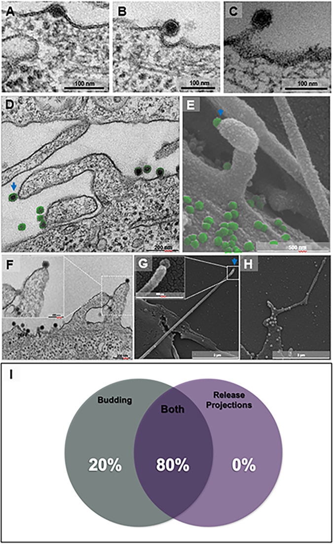
FIG 5.
Chikungunya virus release by cellular protrusions in addition to budding. Vero cells were infected with chikungunya virus (CHIKV) at a multiplicity of infection (MOI) of 5.0 at 8 hpi (A to D and F) by transmission electron microscopy (TEM). Furthermore, for scanning electron microscopy (SEM), cells were infected with CHIKV at an MOI of 5.0 and assessed at 8 hpi (E, G, and H). (A to C) Release of CHIKV by budding, acquiring an early envelope from the cell membrane (A), finishing envelope formation (B), and almost completing release (C). (D to H) Release by cell membrane protrusions. A protrusion with a single CHIKV particle (blue arrow) on the tip and other particles near the cell surface (in green) in a transmission micrograph (D) and a scanning micrograph (E). The transmission micrograph shows two projections with a single CHIKV particle at the tip (F). The scanning micrograph shows a long actin tail with a single particle at the tip (G, blue arrow). The scanning micrograph shows multiple particles over the cell surface and some particles along the tail with a particle at the tip (H). Percentage of presence of release protrusions and budding present in 25 infected cells (I). Electron micrographs are shown with scale bars. Bars, 100 nm (A); 100 nm (B); 100 nm (C); 200 nm (D); 500 nm (E); 200 nm (F); 2 μm (G); 2 μm (H).

