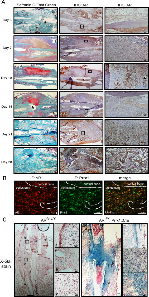
Fig. 1. The AR is expressed in the periosteum during the fracture healing process.
A Histological sections of post-fracture calluses at 3, 7, 10, 14, 21 and 28 days were analyzed by Safranin O/Fast Green staining and immunohistochemical analysis of AR expression. Scale bar = 100 µm. B Double-immunofluorescence staining revealed co-localization of the AR (red) and Prrx1 (green) in the periosteum of post-fracture calluses at 10 days. Scale bar = 50 µm. C X-gal–stained cells were observed in the post-fracture callus at 10 days in AR-/Y;Prrx1::Cre::Rosa26-LacZ mice (right panel); and ARflox/Y mice (left panel) were used as negative controls. Scale bar = 50 µm. a. periosteum; b. mesenchyme; c. chondrocytes. Scale bar = 100 µm.

