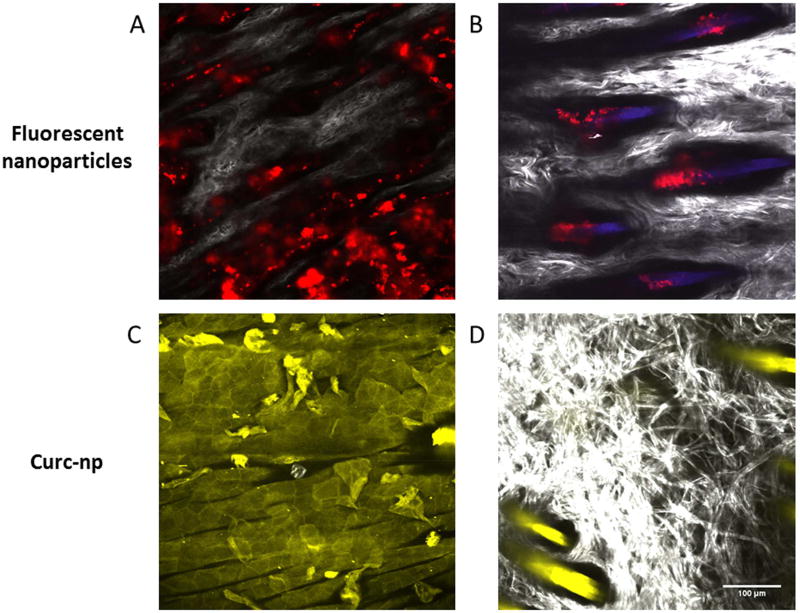
Figure 1. Two-photon microscopy of nanoparticles topically applied to rat abdomen.
(A) Image generated 1-hour post-application of nanoparticles to the shaved abdomen of a ZDF rat. Red fluorescently labeled nanoparticles are seen to reside in skin furrows between the white collagen fibers that make up dermal papillae at a depth of 45µm. (B) Image generated 24-hours post-application of nanoparticles show residence in hair follicles. (C) Image generated 2-hours post-application of curc-np at 12µm depth and (D) 45 µm depth; the yellow color is due to the fluorescence of curcumin.

