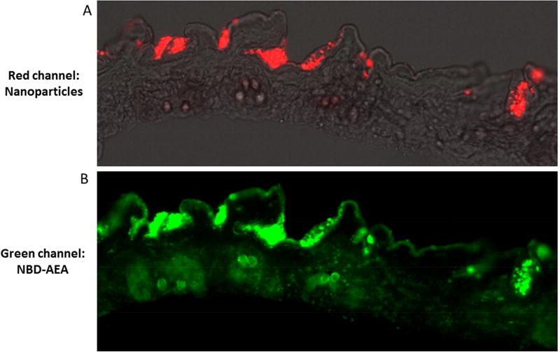
Figure 2. Epifluorescent histologic image of ZDF rat skin treated with fluorescent nanoparticles.
Images generated 1-hour post-application of red labeled nanoparticles loaded with green colored lipip in an 8µm thick slice of ZDF rat abdominal skin. (A) is an overlay of the phase and red channels showing labeled nanoparticles have penetrated the thin epidermis and have begun to collect in hair follicles. (B) is the green channel of the same image showing the labeled lipid diffusing out from nanoparticles across the epidermis as well as deeper at the base of hair follicles.

