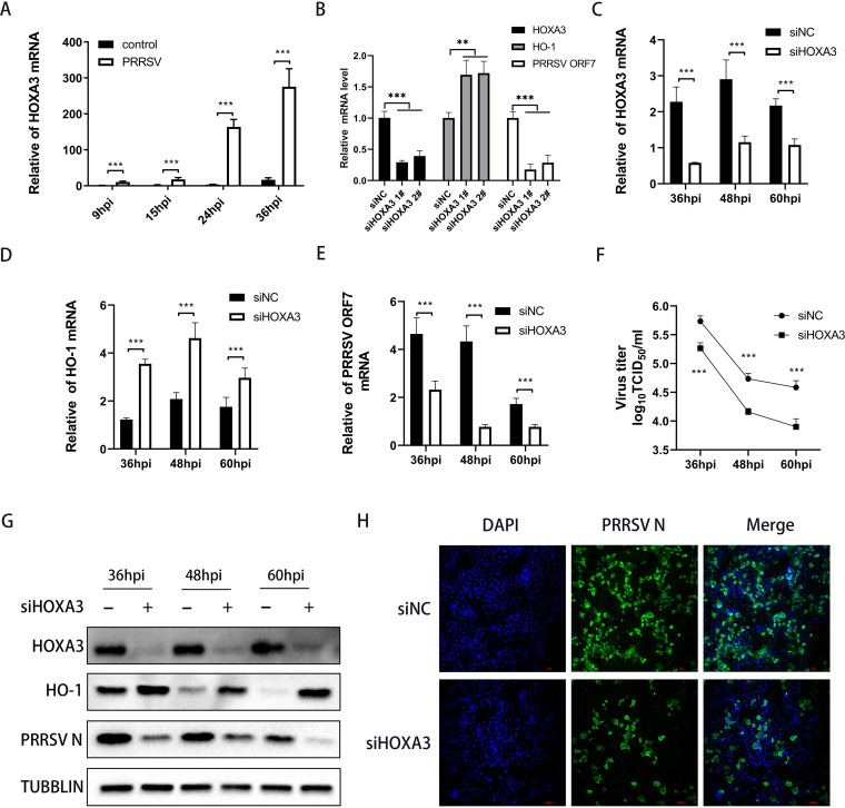
FIG 2.
Knocking down HOXA3 upregulated HO-1 expression and inhibited PRRSV replication. (A) PAMs were infected with PRRSV at an MOI of 0.1. Samples were collected at 9, 15, 24, and 36 hpi; RT-qPCR determined HOXA3 mRNA levels. (B) RT-qPCR analysis of HOXA3, HO-1, and PRRSV ORF7 mRNA in MARC-145 cells transfected with negative control siRNA (siNC) or HOXA3 siRNA (siHOXA3) no. 1 and 2 for 24 h followed by infection with PRRSV for 36 h. (C to E) MARC-145 cells were transfected with siHOXA3 no 1 or siNC of 60 nM for 24 h before infection with PRRSV at an MOI of 0.1. Samples were collected at 36, 48, and 60 hpi. HOXA3 (C), HO-1 (D), and ORF7 (E) mRNA levels were determined by RT-qPCR. (F) Cell culture supernatants were collected at the indicated times. The TCID50 assay was performed to determine the levels of supernatant virus production. (G) Protein levels of HOXA3, HO-1, PRRSV N, and tubulin at the indicated time points were measured by Western blotting. (H) The expression of the N protein was determined by IFA at 36 hpi, with MARC-145 cells transfected with siNC included as a control. Results are expressed as means ± SD of three independent replicates. Statistically significant values were denoted as follows: ***, P < 0.001.

