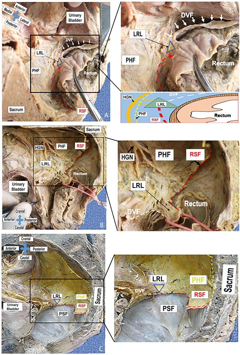
Figure 6.
Bilateral attachment of the posterior and anterior rectal fasciae. (A) Transverse view; (B) lateral view; (C) lateral view (the rectum is resected). White dashes: Denonvilliers’ fascia (DVF); red dashes: the rectosacral (Waldeyer’s) fascia (RSF); yellow dashes: pre-hypogastric fascia (PHF); triangle: lateral rectal ligament (LRL); HGN, hypogastric nerve; PSF, presacral fascia.

