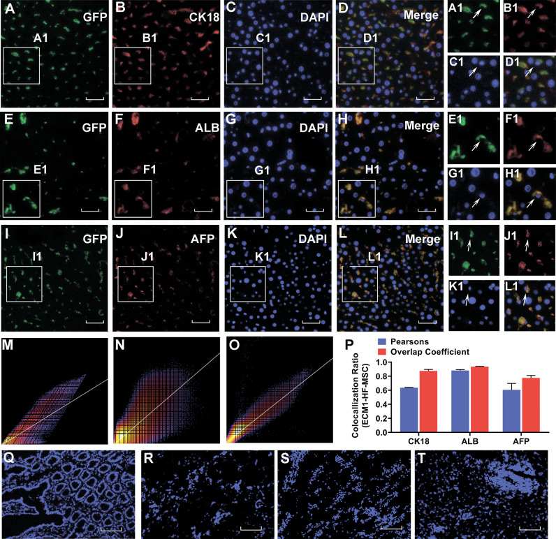
Fig. 6. ECM1-overexpressing HF-MSCs home to the injured liver site and differentiate into hepatocyte-like cells.
Colocalization of GFP-expressing ECM1-HF-MSCs and the hepatic-specific surface markers CK18 A–D, ALB E–H, and AFP I–L. A1–L1 Magnification of regions of (A–L). M–O Scatter plots of GFP-expressing ECM1-HF-MSCs and CK18, ALB, and AFP levels. P Pearson’s correlation and the overlap coefficient of liver sections costained for CK18, ALB, and AFP in the ECM1-HF-MSC group. Q–T GFP-labeled ECM1-HF-MSCs were rarely observed in the intestine, kidney, lung, or spleen. Scale bar (A–L), 50 μm; Scale bar (Q–T), 200 μm.

