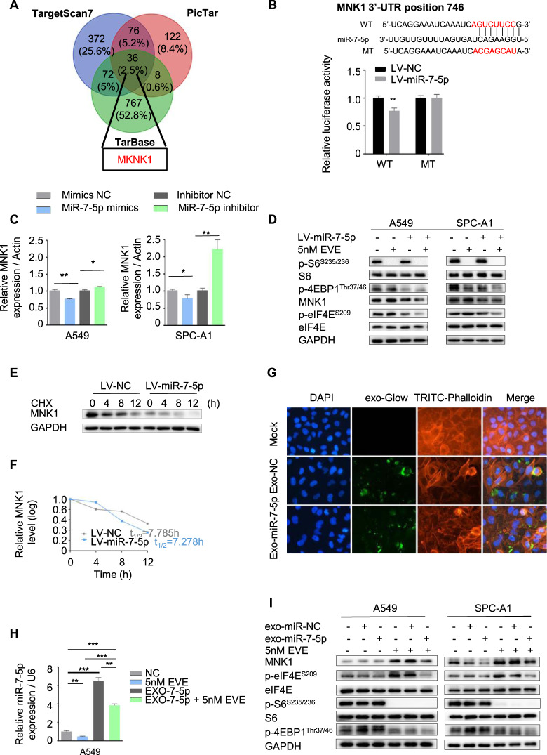
Fig. 2. Loss of intracellular miR-7-5p induced phosphorylation of MNK/eIF4E axis, but supplement of extra exosomal miR-7-5p could reverse it.
A The MKNK1 was the target gene of miR-7-5p predicted by the bioinformatics analysis. B The binding sites of miR-7-5p in 3′-UTR region of MKNK1. Wild-type and mutant sequences were indicated (upper). The LV-miR-7-5p and control A549 cells were transfected with a luciferase reporter containing the 3ʹ-UTR (WT or Mut) of MNK1, indicating MKNK1 was the target gene of miR-7-5p. C Changing the levels of miR-7-5p in A549 and SPC-A1 cells by indicated mimics or inhibitor for 24 h, the mRNA of MKNK1 were determined by qPCR. D The LV-miR-7-5p and control A549 and SPC-A1 cells were treated with DMSO or 5 nM Everolimus alone or in combination for 24 h to detect the expression of MNK1 and p-eIF4ES209 and downstream of mTORC1, including p-S6/S6 S235/236 and p-4EBP1Thr37/46, by Western blotting. E The LV-miR-7-5p A549 cells were treated with cycloheximide (20 μg/mL) and collected in the indicated times. The protein of MNK1 was detected by Western blotting. F The half-life of the MNK1 protein was calculated. G Fluorescently labeled exosomes derived from indicated A549 cells were internalized by A549 cells (exo-NC means exosomes derived from A549 infected with LV-NC; exo-miR-7-5p means exosomes derived from A549 infected with LV-miR-7-5p). Representative images were filmed after cells were fixed and stained (magnification, 400×). H The miR-7-5p level was detected by qPCR in A549 cells treated with Everolimus or miR-7-5p loaded exosomes alone or in combination. I The expression of MNK1 and p-eIF4ES209 and downstream of mTORC1 including p-S6/S6 S235/236 and p-4EBP1Thr37/46, in A549 and SPC-A1 cells treated with Everolimus or miR-7-5p loaded exosomes alone or in combination were detected by Western blotting.

