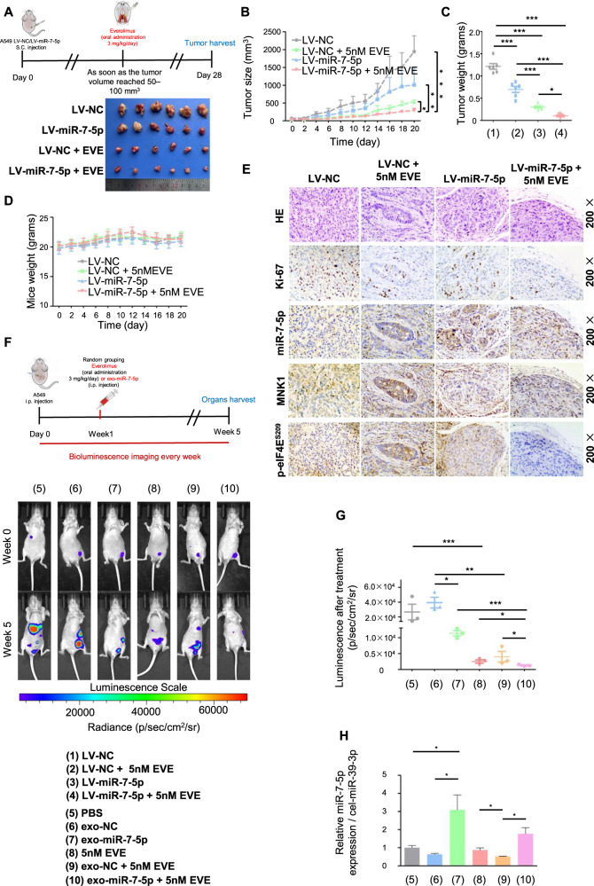
Fig. 5. Exosomal miR-7-5p enhanced the anticancer therapeutic efficacy of Everolimus in vivo.
Five-week-old male nude mice were randomly divided into two groups to establish LV-NC and LV-miR-7-5p A549 cells subcutaneous xenograft nude mice models. As soon as the tumor volume reached 50-100mm3, they would be divided into four groups named as 1) The LV-NC group, 2) The LV-miR-7-5p group, 3) The LV-NC combined Everolimus group and 4) The LV-miR-7-5p combined Everolimus group. A Upper: Schematic illustration of Everolimus oral administration for the subcutaneous xenograft nude mice models. Lower: Comparison of tumor engraftment size and weight in nude mice subcutaneously injected into the flanks with indicated A549 cells and indicated treatment. B The mice xenograft tumor growth curves of the four groups, C the tumor weight and D the body weight. E Immunohistochemical/In situ hybridization staining for the proliferation marker Ki67, MNK1, p-eIF4ES209 and miR-7-5p in the tumor tissues. The A549 cells labeled with luciferase gene were injected into the abdominal cavity of nude mice, and the baseline level of tumor cells was monitored by live imaging technology immediately after injection. One week later, they would be randomly divided into 6 groups for receiving PBS, exo-NC, exo-miR-7-5p, Everolimus, exo-NC combined with Everolimus and exo-miR-7-5p combined with Everolimus treatment, respectively. F Upper: Schematic illustration of abdominal tumor xenograft nude mice models. Lower: Luminescence signals of intraperitoneal A549-luciferase cells with different treatment groups at the indicated week. Results are shown as the mean ± SD. G Analysis of intraperitoneal A549-luciferase cells accepted different treatment groups at the indicated week. H The levels of exo-miR-7-5p from the plasma of nude mice receiving treatments above were measured by qPCR. * P < 0.05, ** P < 0.01, ***P < 0.001.

