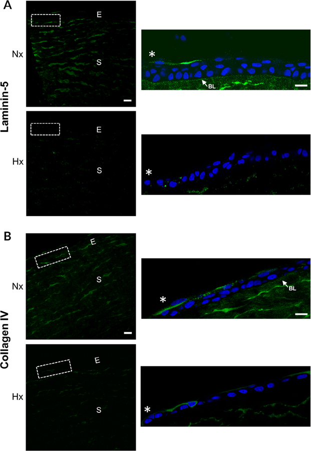
Fig. 3.
Hypoxia modulates the localization of laminin and Type IV collagen in wounded corneas. Epithelial abrasions were performed and corneas incubated in normoxic or hypoxic environments for 18 hours. Corneas were immunostained for laminin-5 and Type IV collagen and counterstained with DAPI. Images were obtained at 40× magnification using a Zeiss Axiovert LSM 700 confocal microscope. The region outlined is shown in inset. A, Laminin-5 is reduced along the basal lamina and in the stroma of hypoxic corneas. Arrow indicates laminin-5 localization to the basal lamina. B, Type IV collagen localization is reduced along the basal lamina and stroma after exposure to hypoxic conditions. Arrow indicates Type IV collagen localization to the basal lamina in the normoxic cornea. Scale bar = 100 μm. *, wound edge. Images are representative of a minimum of three independent experiments. Nx, normoxia; Hx, hypoxia; E, epithelium; S, stroma; BL, basal lamina.

