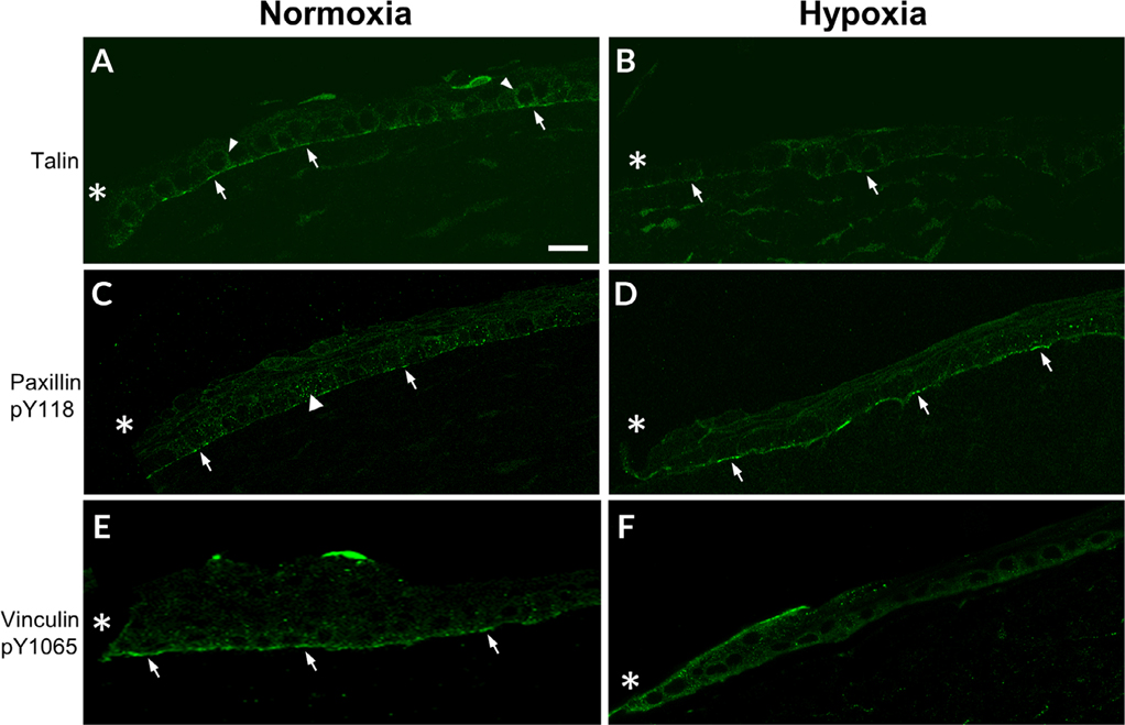
Fig. 4.
Localization of focal adhesion proteins is altered under hypoxic conditions. A–D, Corneas were wounded, incubated in normoxic or hypoxic environments for 18 hr, and stained for focal adhesion proteins. Images were obtained at 40× magnification using a Zeiss Axiovert LSM 700 confocal microscope and are representative of a minimum of three experiments. A, Talin localizes along the basal surface (arrows) and along basolateral surface (arrowheads) of corneal epithelial cells. B, Talin is diminished after exposure to hypoxic conditions. Arrows indicate localization to the basal surface. C, Paxillin pY118 localizes to the basal surface (arrows) and the basolateral surface (arrowhead) of epithelial cells D, Paxillin pY118 localization is present along the basal surface (arrows). E, Vinculin pY1065 localizes to the basal surface (arrows). F, Vinculin pY1065 is present at the wound edge along basal and apical surface. *, wound edge. Scale bar = 50 μm.

