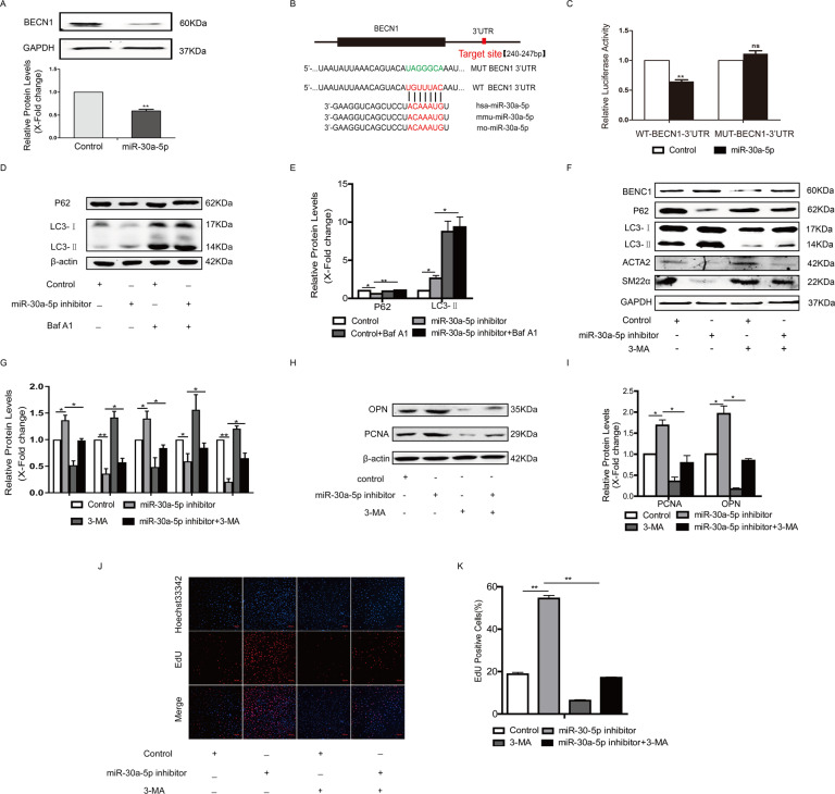
Fig. 4. MiR-30a-5p decreased the expression of beclin1 by targeting the 3’UTR of beclin1.
A HA-VSMCs were transfected with miR-30a-5p mimics, and then the expression of beclin1 was detected by western blotting (**P < 0.01, n = 3). B Alignment of the miR-30a-5p “seed region” with beclin1 3′-UTRs among different species. C HA-VSMCs were transfected with beclin1 3′-UTR or mutant beclin1 3′-UTR plasmids together with miR-30a-5p mimic or control for 24 h, followed by luciferase assays (**P < 0.01, n = 3). D and E HA-VSMCs were transfected with miR-30a-5p inhibitor or control for 24 h and then treated with bafilomycin A1 for 2 h. Cells were harvested and accumulation of P62 and LC3 was measured by western blotting (*P < 0.05, **P < 0.01, n = 3). F–I HA-VSMCs were transfected with miR-30a-5p inhibitor for 4 h and then treated with 5 mM 3-MA for another 48 h. Western blot images and quantification in each group are shown (*P < 0.05, **P < 0.01, n = 3). J and K Cellular proliferation was evaluated by EdU staining (**P < 0.01, n = 3).

