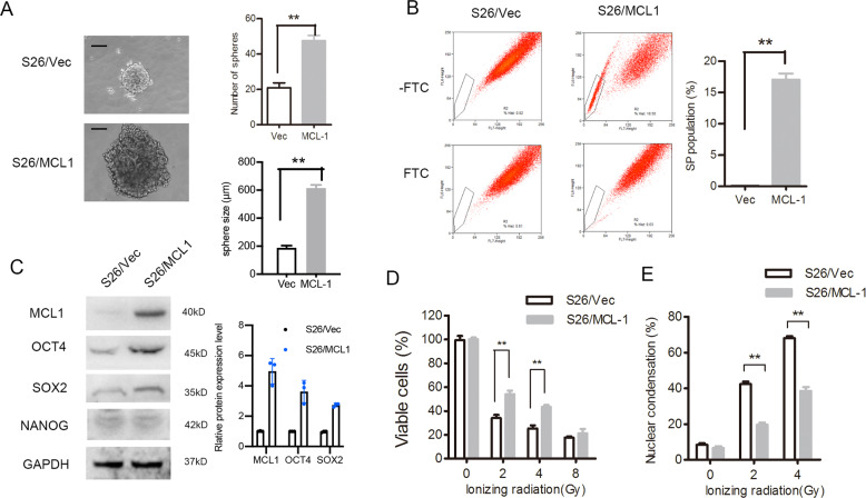
Fig. 4. Expression of MCL-1 in NPC cells induced CSC characteristic and radiation resistance.
S26 cells stably overexpressing MCL-1 or empty vector (Vec) were analyzed as follows. A The representative images of single-cell suspensions are shown (left panel) and spheroids were counted (right upper panel), and the size were measured (right lower panel), **p < 0.01, Student’s t-test. B Percentages of SP cells are shown in the left panel, and the right panels compare the populations of SP, n = 3, *p < 0.05, Student’s t-test. C Protein levels of stem-cell markers were determined by western blotting, GAPDH was used as the loading control. D Cell viability after treatment with ionizing radiation determined by MTS assay, **p < 0.01, Student’s t-test. E Qualification of nuclear condensation after treatment with different doses of radiation are shown, **p < 0.01, Student’s t-test. (NPC, nasopharyngeal carcinoma; CSC, cancer stem cell; MTS, 3-(4,5-dimethylthiazol-2-yl)-5-(3-carboxymethoxyphenyl)-2-(4-sulfophenyl)-2H-tetrazolium; SP, side population).

