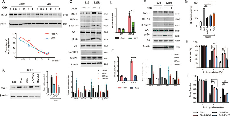
Fig. 5. ROS-AKT feedback loop activation regulates the protein synthesis of MCL-1.
A S26 and S26-R cells were treated with CHX (50 μg/mL), lysates were collected at the indicated times. (Upper) Immunoblotting analysis of MCL-1 levels. (Lower) MCL-1 levels were quantified and normalized to the signal of β-actin. B S26-R and S26 cells were treated with CHX (50 μg/mL) or combined with MG132 (5 μM) for 2 h. Cell lysates were subjected to western blotting. β-actin was used as the loading control. C, D S26-R and S26 cells were treated with the AKT inhibitor MK2206 (1 μM) for 2 h, and then cell lysates were subjected to immunoblotting (C), β-actin was used as the loading control, and quantification of ROS levels is shown (D) (n = 3); *p < 0.05, **p < 0.01 compared with the 0 h control. E, F S26 and S26-R cells were treated with N-acetylcysteine (NAC, 5 μM) for 2 h, E Quantification of ROS levels measured by DCFH-DA is shown (n = 3; *p < 0.05, **p < 0.01 compared with the 0 h control); F cell lysates were subjected to immunoblotting, β-actin was used as the loading control. G S26-R cells and s26 cells were treated with the AKT inhibitor or NAC, then the cells were cultured for 10 days, and the formed spheroids were counted and compared, **p < 0.01, *p < 0.05, Student’s t-test. H, I S26-R cells and S26 cells were treated with the AKT inhibitor or NAC for 2 h, and then cells received the indicated dose of irradiation, and cell viability was determined by the MTS assay 2 days post-irradiation, and clony formation was determined by stained with methylene blue and counted 10 days post-irradiation, **p < 0.01, *p < 0.05, Student’s t-test. (CHX, cycloheximide; ROS, reactive oxygen species; DCFH-DA, dichlorofluorescin-diacetate; MTS, 3-(4,5-dimethylthiazol-2-yl)-5-(3-carboxymethoxyphenyl)-2-(4-sulfophenyl)-2H-tetrazolium).

