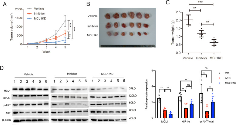
Fig. 6. Inhibition of AKT-MCL-1 signaling increased antitumor effect of radiation therapy in vivo.
S26-R cells transfected with MCL-1 shRNA(MCL1/kd) or negative control shRNA were injected subcutaneously into BALB/c nude mice. Then, S26-R cells transfected with the control scrambled shRNA were treated with AKT inhibitor (MK2206, AKTi) or vehicle (veh, used as control) during treatment with radiation. A Tumor volume growth curves; data are shown as the mean ± SD. (**p < 0.01, ***p < 0.001, Student’s t-test). B Images of tumors from all mice in the indicated groups (n = 6/group). C Weight of tumors formed in the indicated groups; data are shown as the mean ± SD. (**p < 0.01, ***p < 0.001, Student’s t-test). D Tumor lysates were subjected to immunoblotting, β-actin was used as the loading control (left panel). Levels of the indicated proteins were quantified and normalized to the signal of β-actin, and phospho-AKT were normalized total AKT and β-actin (right panel).

