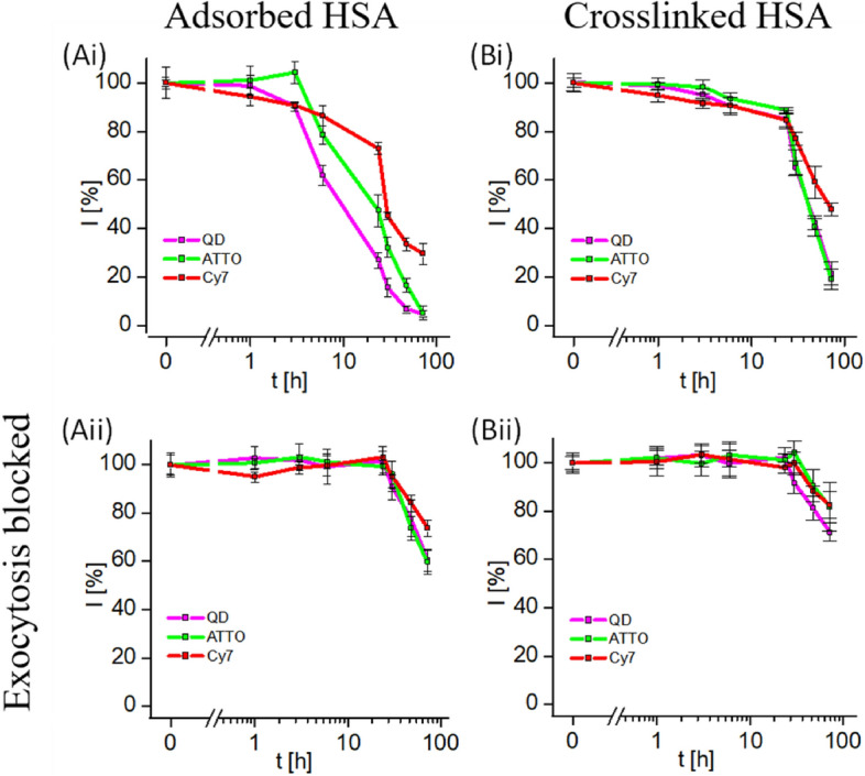
Fig. 5.

Three different compartments of quantum dots were fluorescence labeled by different colors: quantum dots (QDs) with their intrinsic fluorescence, the polymer surface coating with the organic fluorophore ATTO (ATTO488; ATTO-TEC GmbH, #AD488-91) [36], and human serum albumin as model protein with the organic fluorophore Cy7 (Sulfo-Cyanine7 NHS ester, Lumiprobe, #25320). The reduction of intracellular fluorescence after free NPs in the extracellular medium around cells with endocytosed NPs had been removed by rinsing. Image taken with permission from Carrillo-Carrion et al. [36]. These data were recorded with HeLa cells and CdSe/ZnS NPs with a core diameter of around 5.5 nm, coated with the polymer poly(isobutylene-alt-maleic anhydride)-graft-dodecyl (PMA), which was fluorescence labeled with ATTO
