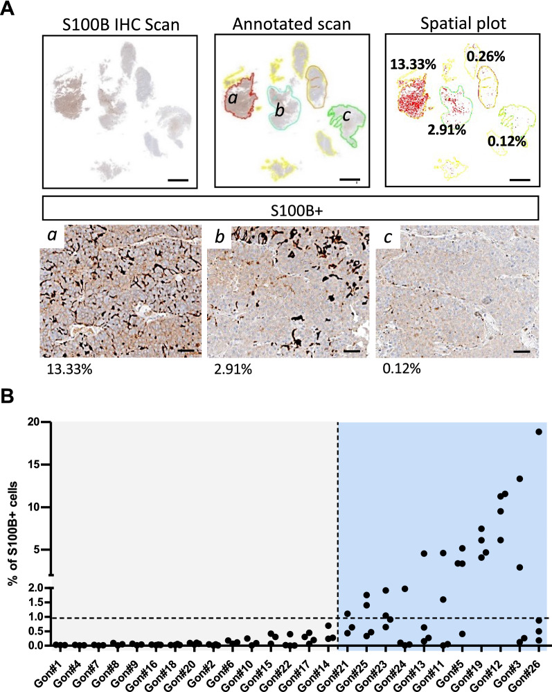
Fig. 4.
S100B + cells show an interpatient and an intratumoural heterogeneous distribution in gonadotroph tumours. A Quantification of S100B + cells on a gonadotroph tumour (Gon#3) showing a variation of more than 100-fold between different areas quantified. Top panel shows the four tumour areas (colour-coded in red, blue, orange, and green) that we defined and quantified on the scanned slide. The spatial plot represents each quantified cell as a red dot, and percentages for each quantified areas are noted. Lower panel shows enlarged views of S100B immunostaining in the indicated area (a, b, and c). Scale bars = 2 mm (top panel), 50 µm (lower panel). B Graph showing the percentage of S100B + cells quantified in multiple areas of 26 gonadotroph tumours (Gon#). Each dot plots the percentage of S100B + cells in a defined tumour area. Horizontal dashed line corresponds to a threshold of 1% S100B + cells. Vertical dashed line splits gonadotroph tumours with almost no expression of S100B among quantified areas and gonadotroph tumours presenting areas with variable percentages of detected S100B + cells. Abbreviation: immunohistochemistry (IHC)

