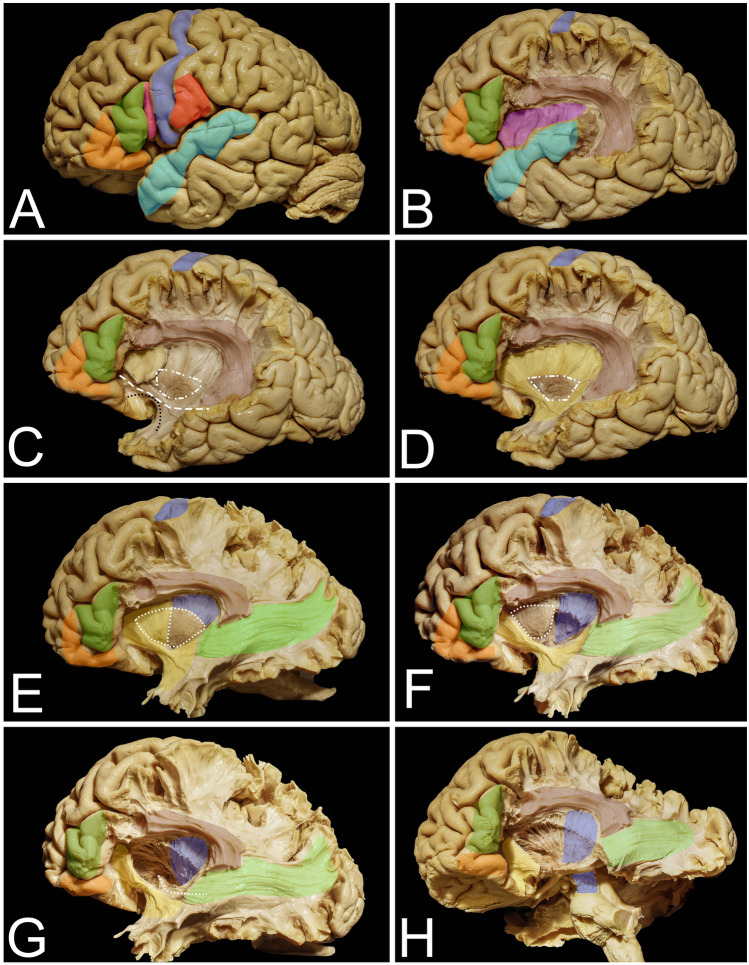
Fig. 1.
The anatomy of cortical and subcortical structures around and on the mesial surface of the insula. A The figure represents the eloquent cortical opercula that surround the insula. Within the IFG, the pars orbitalis (orange), pars triangularis (green), and pars opercularis (pink) within the dominant hemisphere are related mainly to language function. The posterior precentral gyrus (blue) and postcentral gyrus (red) represent the primary motor and sensory cortex, respectively. The superior temporal gyrus (light blue) represents the temporal operculum, and its posterior part represents the so-called Wernicke’s speech area. B Resection of the posterior part of the frontal operculum and the parietal operculum exposes the AF/SLF (light burgundy) complex, which is located around the insula in most superficial association fiber systems. In addition, the insular cortex (purple) is exposed. C Resection of the insular cortex exposes the extreme capsule (light yellow), which is separated by the claustrum (white square-rectangle line) from the external capsule, which in its anterior and inferior aspect within the limen insulae is composed of UF (black dots) and IFOF (white rectangles) fibers. D Resection of the extreme capsule provides exposure of the external capsule (darker yellow), and the claustrum was left in place. E With resection of the external capsule and claustrum, the putamen (white dots) is exposed (though it is still anteriorly covered by the external capsule); the corona radiata is located above it and mesial to the AF/SLF complex, representing the corticospinal fibers (blue). Extending resection posteriorly beyond the AF/SLF complex reveals the fibers of the sagittal stratum (light green). F Resection of the posterior half of the putamen exposes the fibers of the posterior limb of the internal capsule, which are located deeper than the corona radiata, on the mesial border. G Resection of the anterior half of the putamen exposes the anterior perforated substances from the superior region, and the anterior commissure at depth is visible (white squares). H Resection of the temporal lobe reveals the corticospinal fibers within the posterior limb of the internal capsule as well as the fibers within the cerebral peduncle

