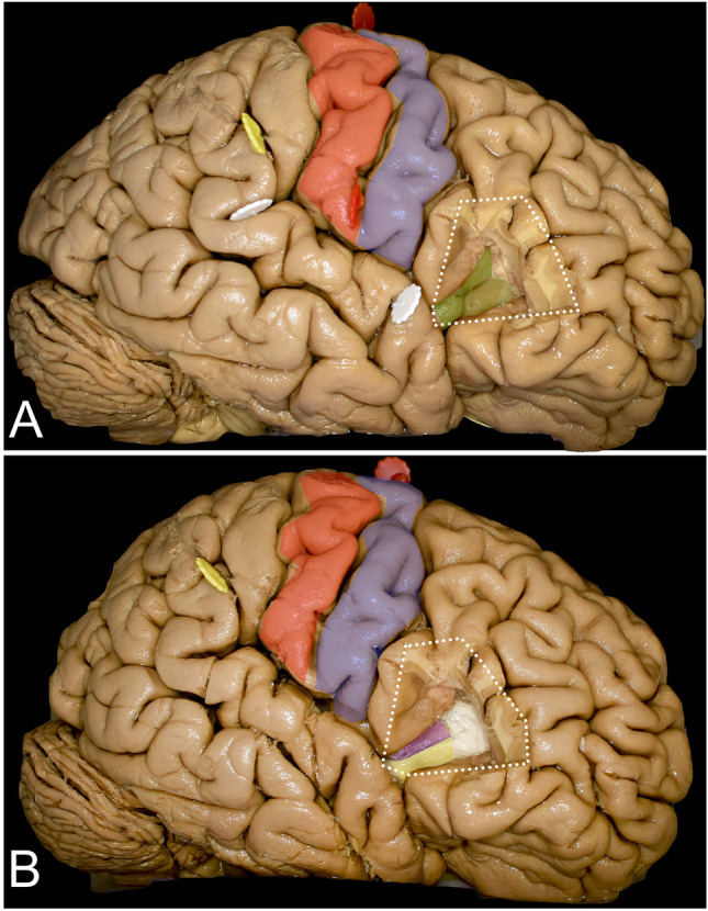
Fig. 5.

Exposure of the insular cortex and subcortical region was achieved with frontal operculum removal. A Resection of the pars triangularis (white dots) of the IFG provided exposure of the superior aspects of the anterior (light green) and anterior half of the middle short (darker green) gyrus. B On the subcortical level, this area corresponds to the frontal horn of the lateral ventricle (white) and below to the putamen (purple) posteriorly and the external capsule (yellow) anteriorly
