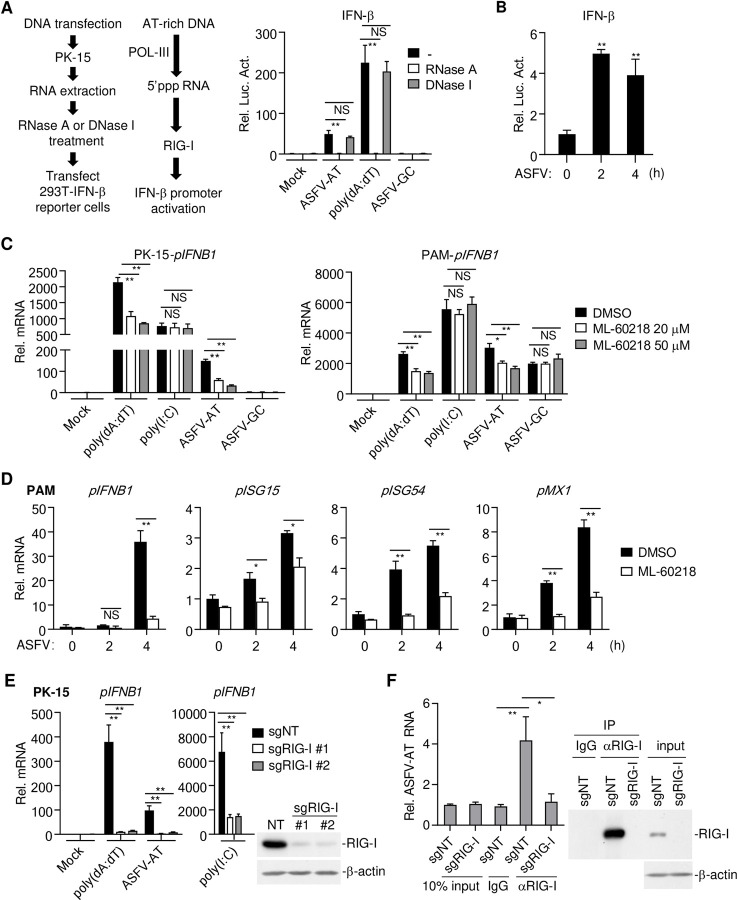
Fig 2. AT-rich regions of the ASFV genome activate RNA Pol-III-RIG-I axis.
(A) RNAs extracted from poly(dA:dT)-, ASFV-AT-, ASFV-GC (1 μg/ml) or mock-transfected PK-15 cells (2×10⁶) were treated with RNase A or DNase I at 37°C for 1 hour. The RNAs were then transfected into HEK293T (2 μg/ml) that transfected with IFN-β-luciferase reporter. Six-hours later, luciferase assays were performed. The flow diagram of experiment was shown on the left. ASFV-AT indicates ASFV-AT-#1 hereafter. (B) PAMs (2×10⁶) were mock infected or infected with ASFV (MOI = 0.1) for the indicated times. RNAs were extracted and treated with DNase (200 U/ml/mg RNA) at 37°C for 1 hour. The RNAs were then transfected into HEK293T (2 μg/ml) that transfected with IFN-β-luciferase reporter. Six-hours later, luciferase assays were performed. (C) PK-15 cells or PAMs (1×10⁶) were pre-treated with the indicated concentrations of the RNA Pol-III inhibitor ML-60218 for 6 hours before transfection with poly(dA:dT), ASFV-AT, ASFV-GC or poly(I:C) (1 μg/ml) for 4 hours. The mRNA levels of IFNB1 were analyzed by RT-qPCR. (D) PAMs (2×10⁶) were pre-treated with ML-60218 (50 μM) for 6 hours before infection with ASFV (MOI = 0.1) for the indicated times. The mRNA levels of IFNB1, ISG15, ISG54, and MX1 were analyzed by RT-qPCR. (E) The effects of RIG-I-deficiency on poly(dA:dT)-, ASFV-AT- or poly(I:C)-stimulated IFN-β induction. Two sgRNA were used to knockdown RIG-I in PK-15 cells using CRISPR/Cas9 method (sgNT, non-targeting sgRNA). The mRNA levels of IFNB1 were analyzed by RT-qPCR after the cells were stimulated with transfected DNA or RNA analogs poly(I:C) for 6 hours. The knockdown efficiency of RIG-I was confirmed by western blot. (F) PK-15 cells (2×10⁷) prepared as in (E) were transfected with ASFV-AT-#2 for 3 hours. RIP experiments were performed using the RIG-I antibody, and specific primers were used to detect the enrichment of ASFV-AT by RIG-I in cells. The expression of RIG-I in lysates and immunoprecipitations was detected by western blot analysis.

