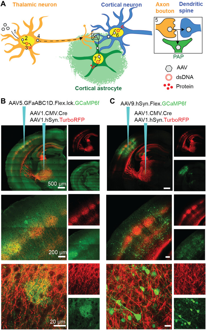
Fig. 1. AAV1 transfer from VPM thalamocortical neurons to BX astrocytes and neurons.
(A) Anterograde AAV transfer hypothesis: (1) AAVs enter neurons at the injection site by endocytosis. (2) Some AAV particles enter the nucleus and (3) release their single-stranded DNA (ssDNA), which is converted to double-stranded DNA (dsDNA) concatemers. The transgene is transcribed and then translated into proteins. (4) A small number of AAV particles are transported anterogradely along the axon to the terminals where (5) they are released and enter adjacent postsynaptic dendrites and perisynaptic astrocytic processes (PAP). Then, they enter the nuclei of (6) neurons and (7) astrocytes and release their ssDNA, which eventually leads to protein synthesis. (B) Intersectional AAV injection strategy for Cre-dependent GCaMP6f (green) labeling of BX astrocytes and TurboRFP (red) labeling of VPM neurons (red), 3 weeks after injection. (C) Intersectional AAV injection strategy for Cre-dependent GCaMP6f (green) labeling of BX neurons and TurboRFP (red) labeling of VPM neurons (red), 2 weeks after injection.

