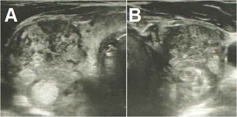
Figure 1.

Thyroid ultrasonography shows increased dimensions of the gland and heterogeneous echotexture, with poorly defined regions of decreased echogenicity and pseudonodules consistent with thyroiditis. There are also multiple hyperechoic nodules in both the right (a) and the left (b) sides.
