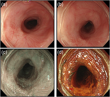
FIGURE 1.

(a) Early esophageal cancer, measuring 10 mm in diameter, in the middle thoracic esophagus. (b) The lesion is located at the site of the post‐endoscopic submucosal dissection (ESD) stricture with a slight distal extension. (c) Narrow‐band imaging of the lesion. (d) Lugol staining of the lesion
