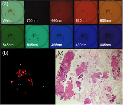
FIGURE 4.

AMUS findings of a specimen of pancreatic adenocarcinoma in the no‐isolation group. (a) The multiband images obtained by the AMUS are shown. (b) The core tissue regions detected using the segmentation algorithm are shown in white. (c) The histological presentation is shown on the bottom right image (40 × magnification). The SVWC length of a specimen of pancreatic adenocarcinoma in the No‐isolation group was 12 mm, whitish core amount of 15 mm2, histological adequacy score of 5, tumor cell content ratio score of 2, and degree of blood contamination score of 3. AMUS, automated multiband imaging system
