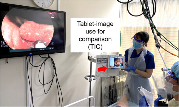
FIGURE 1.

The tablet‐image comparison (TIC). Images obtained via the first modality are displayed on the tablet held next to the monitor by an assistant. According to this, the endoscopist can adjust the amount of air and the angle and distance of the lesion similarly in the second modality and produce images for the comparison of the two modalities under conditions that are largely the same
