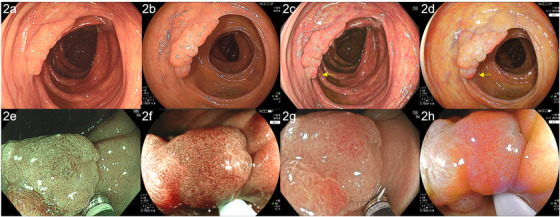
FIGURE 2.

Images of high‐grade dysplasia were obtained by color enhancement imaging (TXI) and linked color imaging (LCI) using the EVIS X1 (CF‐EZ1500DI) and ELUXEO systems, compared by tablet‐image comparison (TIC). (a) A superficial elevated lesion of 30 mm with white light imaging (WLI) using CF‐EZ1500DI (high‐grade adenoma, ascending colon). (b) In WLI performed using the ELUXEO system, comparison by TIC revealed that the redness and brightness of the image were not so different from the redness and brightness on the images obtained by the EVIS X1 system. (c) On TXI, the granules of the lesion were generally reddish, and the redness was particularly emphasized at the depression (yellow arrow). A hyperplastic polyp of 2 mm could be seen (white arrow). (d) Compared to TXI, LCI suppressed the redness overall. The depressed area was more reddish in comparison to the surrounding areas (yellow arrow). Hyperplastic polyps of 2 mm could be clearly seen (white arrow). (e) Magnified narrow band imaging (NBI) clearly showed an irregular surface pattern. The vessel pattern was thin and complex. (f) An image of the same area was more brownish under magnified blue light imaging (BLI) and the surface pattern was less irregular and more enhanced in comparison to images obtained by the EVIS X1 system. (g) TXI magnified observation showed that the surface pattern was irregular and complex, similar to NBI. (h) LCI magnified observation showed that the surface pattern was irregular, similar to BLI
