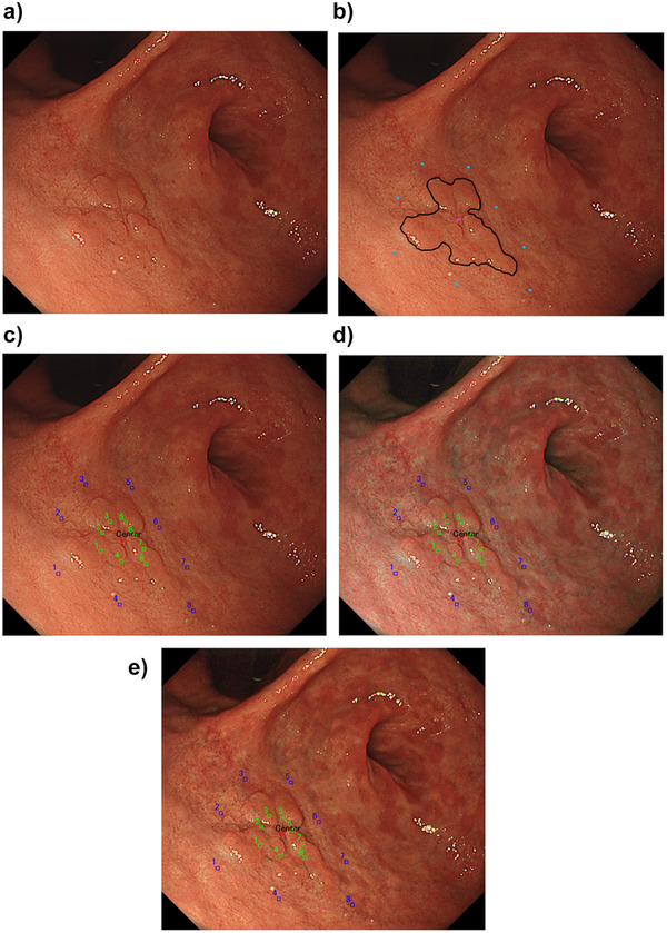
FIGURE 2.

Annotation of lateral margin, peripheral, and inner points on EGC images for color difference analysis. (a) A reddish elevated lesion is seen in the anterior wall of the antrum. (b) An expert endoscopist defined the region of interest and delineated the margin of EGC on the WLI image. The endoscopist manually annotated the center of the lesion, eight equal peripheral non‐cancerous points 5 mm outside the lesion (proximal, distal, anterior, posterior, and four midpoints between the two points). (c) Eight innerpoints (green spots) were annotated two‐thirds of the distance from peripheral points to the EGC lesion center. Blue spots indicate the peripheral points. (d) The peripheral and inner points were similarly annotated on the image of TXI mode 1 with the same angle, distance, and air insufflation. (e) The peripheral and inner points were similarly annotated on the image of TXI mode 2 with the same angle, distance, and air insufflation
