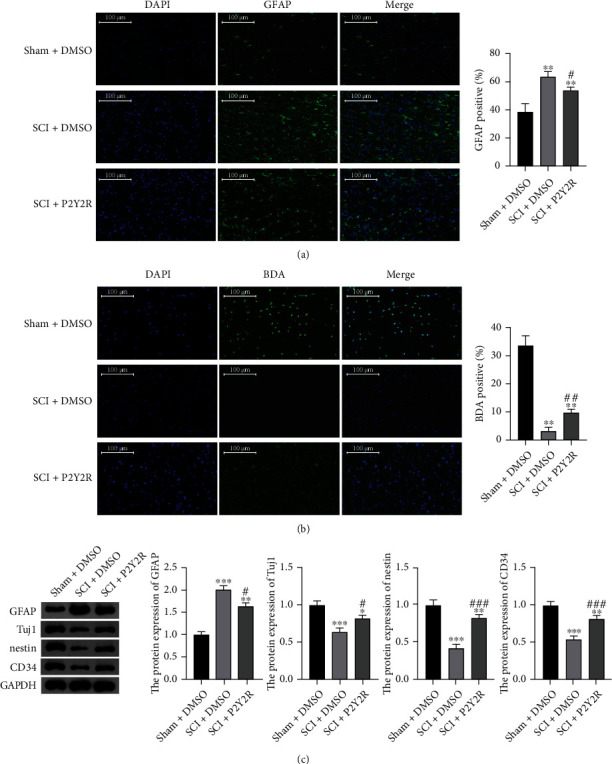
Figure 5.

(a) The expression levels of GFAP detected by immunofluorescence staining. (b) Results of BDA staining. Scale bar = 100 μm. (c) The relative protein expression of GFAP, Tuj1, nestin, and CD34 measured by western blotting. ∗∗p < 0.01 compared with sham+DMSO group; #p < 0.05 and ##p < 0.01 compared with the SCI+DMSO group.
