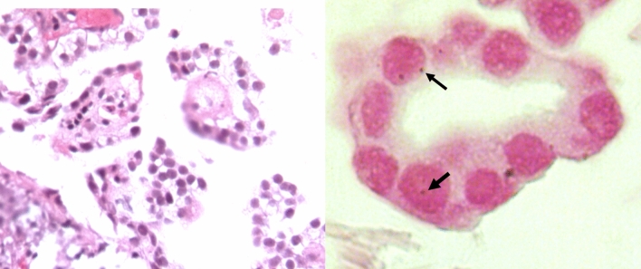
Figure 2.
Representative micrographs of pulmonary adenocarcinoma and its in-situ PCR detection of mycobacterial DNA. Left figure shows well differentiated adenocarcinoma stained with haematoxylin/eosin, neoplastic cells show big hyperchromatic basophilic nucleus. The right figure shows high power magnification of neoplastic gland, some cells show positivity for mycobacterial DNA (IS-6110) detected by in situ RT-PCR (dark blue dots, arrows) in the nucleus stained by nuclear fast red.

