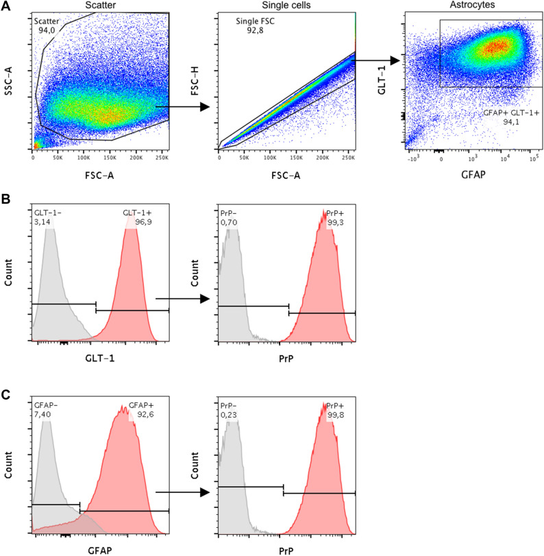
Figure 1.
Composition of primary bank vole glia cells at the time of infection (20 days post isolation, passage 2). Characterisation was performed by flow cytometry analysis based on expression of GFAP and GLT-1, two specific markers for cells of astrocytic phenotype. Additionally, levels of PrPC expression were determined as crucial factor for prion propagation. (A) Proportion of the analysed cells, which were found to co-express GFAP and GLT-1. Cell debris and doublets were excluded from the analysis by gating as illustrated. (B) Histogram of those cells, which were stained for GLT-1 and (C) for GFAP. PrPC expression of each subpopulation is shown in the respective second panel. Red areas of the histograms refer to stained samples and grey areas to the corresponding control incubated without the target antibody. The experiment was carried out in triplicate, of which one representative is shown. According to the analysis the glia cell culture mainly consisted of astrocytes with high PrPC expression, which provided the basis for an infectible cell assay. FSC: forward scatter, SSC: side scatter.

