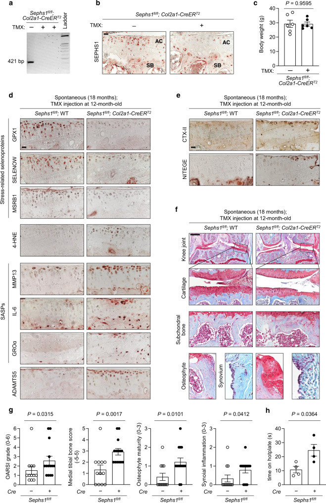
Fig. 3. Chondrocyte-specific Sephs1 knockout in adult mice accelerates aging-associated OA development in knee joints.
a PCR verification of Sephs1 inducible conditional knockout (iCKO) after five intraperitoneal injections of tamoxifen (TMX) in 12-week-old Sephs1fl/fl; Col2a1-CreERT2 mice. b Immunostaining of SEPHS1 in knee joint sections displaying the articular cartilage (AC) and subchondral bone (SB) of 21-week-old WT and Sephs1-iCKO littermates. c Body weight measurements at 8 weeks after five times injections of vehicle or TMX in 12-week-old Sephs1fl/fl; Col2a1-CreERT2 mice (n = 6). d, e Sephs1fl/fl or Sephs1fl/fl; Col2a1-CreERT2 mice were injected with TMX at 12 months of age, and the appearance of aging-associated OA phenotypes was analyzed at 18 months. d Stress-related selenoproteins (GPX1, SELENOW, and MSRB1), 4-HNE, SASPs (MMP13, IL-6, and GROα), ADAMTS5, and e cartilage matrix neoepitopes (telopeptides of type II collagen, CTX-II and aggrecan neoepitope, NITEGE) were detected by immunohistochemistry in cartilage sections. f Joint sections were stained with safranin O, fast green, and hematoxylin. The inset in the images is shown as magnified images in the bottom row. g Scores of OA manifestations, including cartilage destruction, subchondral bone sclerosis, osteophyte formation, and synovial inflammation (n = 12 for Sephs1fl/fl; n = 14 for Sephs1fl/fl; Col2a1-CreERT2). h Hotplate pain assay in 18-month-old WT and Sephs1-iCKO mice (n = 4). Scale bars: b, d, e 25 μm, f 500 μm. c, g, h Data represent means ± s.e.m. P values are from two-tailed t test (c, h) or two-tailed Mann–Whitney U test (g). Cohen’s d effect sizes are provided in Supplementary Table 8. Mankin scores and SBP thickness measurements are provided in Supplementary Figs. 11 and 12.

