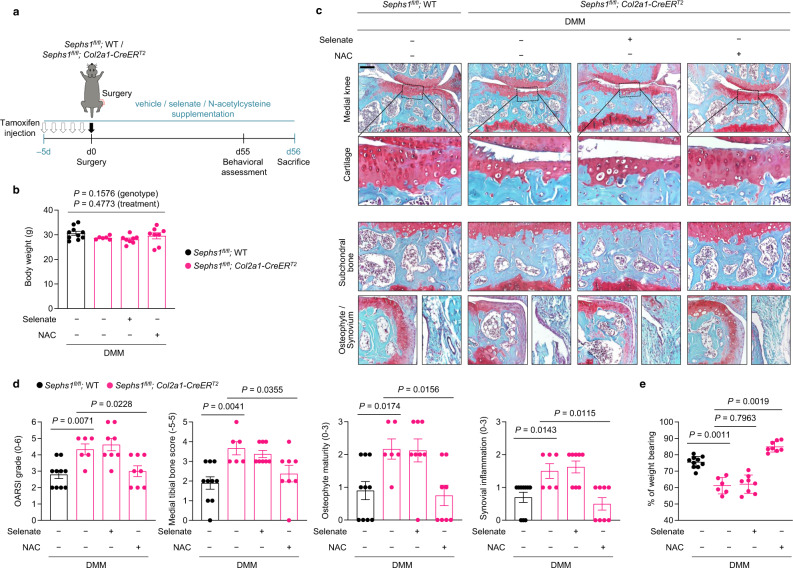
Fig. 5. NAC treatment rescues the exacerbated OA phenotypes in Sephs1-iCKO mice.
a Schematic illustration of NAC or dietary selenate supplementation in the post-traumatic OA model of Sephs1-iCKO mice. b Body weight of 21-week-old DMM-operated mice after completion of the supplementation scheme (n = 10 for DMM-operated WT mice treated with vehicle; n = 6 for DMM-operated Sephs1-iCKO mice treated with vehicle; n = 8 for DMM-operated Sephs1-iCKO mice supplemented with selenate; n = 8 for DMM-operated Sephs1-iCKO mice treated with NAC). c Joint sections were stained with safranin O, fast green, and hematoxylin. The inset in the images is shown as magnified images in the bottom row. d Cartilage destruction, subchondral bone sclerosis, osteophyte formation, and synovial inflammation determined by safranin O/hematoxylin staining and scored (n = 10, 6, 8, 8 respectively). e The percentage of weight placed on the DMM-operated limb versus the contralateral limb of WT and Sephs1-iCKO mice treated with or without NAC or selenate determined using a static weight bearing test (n = 10, 6, 8, 8 respectively). Scale bar: c 200 μm. b, d, e Data represent means ± s.e.m. P values are from two-way ANOVA followed by Tukey’s post hoc test (b) or S–R–H test followed by Mann–Whitney U test (d, e). Cohen’s d effect sizes are provided in Supplementary Table 8. Mankin scores and SBP thickness measurements are provided in Supplementary Figs. 11 and 12.

