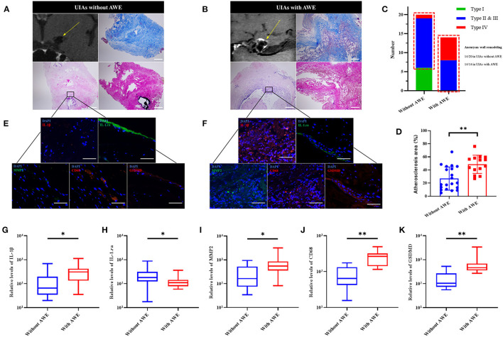
Figure 3.
The GSDMD and the inflammatory factors in UIAs with and without AWE. (A,B) Two representative cases of histological analysis for UIAs with and without AWE. The H-E staining indicated a severe wall remodeling and inflammation infiltration in the UIAs with AWE. The Masson staining showed that atherosclerosis was more significantly changed in the UIAs with AWE. The EVG staining reported the reduced elastic fibers in the UIAs with and without AWE. The scale bar corresponded to 100 μm. (C) The histograms of aneurysm wall remodeling between UIAs with and without AWE. Thus, it was known that all UIAs with AWE were manifested as significant wall remodeling (type II & III, or type IV). The red box indicated the number of aneurysm wall remodeling. (D) The atherosclerosis area between UIAs with and without AWE. The UIAs with AWE had a larger atherosclerosis area compared with UIAs without AWE. (E,F) The IL-1β, IL-1.ra, GSDMD, CD68 and MMP2 in UIA tissues were detected in this study. The scale bar corresponding to 40 μm. (G–K) The box plot of the relative levels of IL-1β, IL-1.ra, MMP2, CD68 and GSDMD for UIAs with and without AWE. *P < 0.05; **P < 0.01. UIA, unruptured intracranial aneurysm; AWE, aneurysm wall enhancement; GSDMD, gasdermin D; MMP2, matrix metalloproteinase-2; IL-1β, interleukin 1β; IL-1.ra, interleukin 1.ra.

