FIGURE 1.
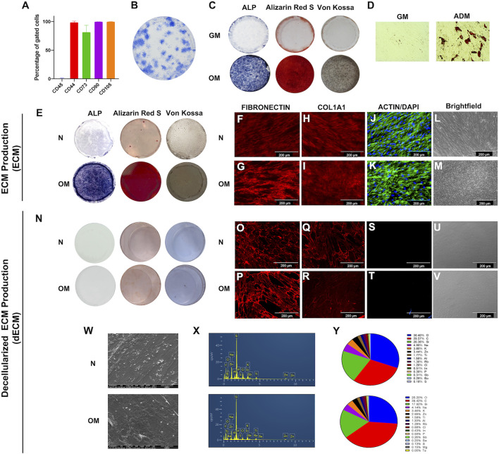
The characterizations of human dental pulp stem cells (DPSCs) and extracellular matrix derived from DPSCs. (A) Stem cell surface markers were evaluated by flow cytometry. (B) The colony-forming unit ability was examined on day 14 using Coomassie blue staining. Osteogenic and adipogenic differentiation ability were determined at days 14 and 16, respectively. (C) ALP activity, calcium and phosphate accumulation at day 14 by staining of BCIP/NBT, Alizarin Red S, and Von Kossa, respectively. (D) The intracellular lipid droplets were determined by using oil red O staining. ECM production from DPSCs were investigated. (E) ALP activity, calcium nodule formation, and phosphate accumulation were examined using BCIP/NBT, Alizarin Red S, and Von Kossa staining, respectively. (F–I) Immunofluorescent staining technique used to determine proteins expression of ECM protein including fibronectin and type I collagen. (J and K) Intracellular cytoskeleton and nucleus was observed using phalloidin and DAPI. (L and M) Brightfield microscope observations of ECM production. Decellularized ECM (dECM) were characterized. (N) ALP and mineral deposition were examined using of BCIP/NBT, Alizarin Red S, and Von Kossa staining, respectively. (O–R) fibronectin and type I collagen were detected using immunofluorescence staining. (S and T) Actin filament and DAPI was examined. (U and V) Brightfield microscope observations of dECM production. (W) Cell morphology was observed using scanning electron microscope. (X and Y) The chemical composition was determined using energy dispersive X-ray analysis, and the percentage of chemical composition was illustrated.

