
Figure 10.
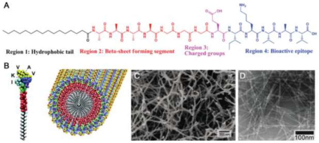
Structure and assembly of PAs: (A) typical structure of a PA, including a hydrophobic tail, β-sheet forming region, charged region, and a bioactive epitope; (B) rendering of a PA with an IKVAV bioactive epitope and the assembly of the IKVAV-presenting PA into nanofibers; (C) scanning electron micrograph of the nanofiber network formed by addition of cell media to an aqueous solution of IKVAV-PA; (D) transmission electron micrograph of the IKVAV-PA nanofiber network. Adapted with permission from Cui et al. [167], Copyright (2010) Wiley-VCH GmbH & Weinheim.