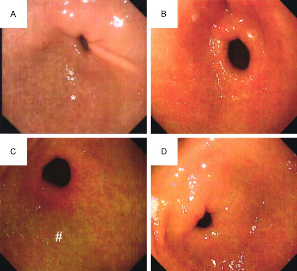
Figure 4.

Typical pictures of gastroscopy. Note: (A) Indicated gastroscopy picture of the experimental group before treatment, while (B) Indicated gastroscopy picture of the experimental group after treatment. (C) Indicated gastroscopy picture of the control group before treatment, while (D) Indicated a gastroscopy picture of the control group after treatment. * Mucosa atrophy and submucosal vascular penetration, # The surface of mucous membrane is rough and uneven, and erosion is visible.
