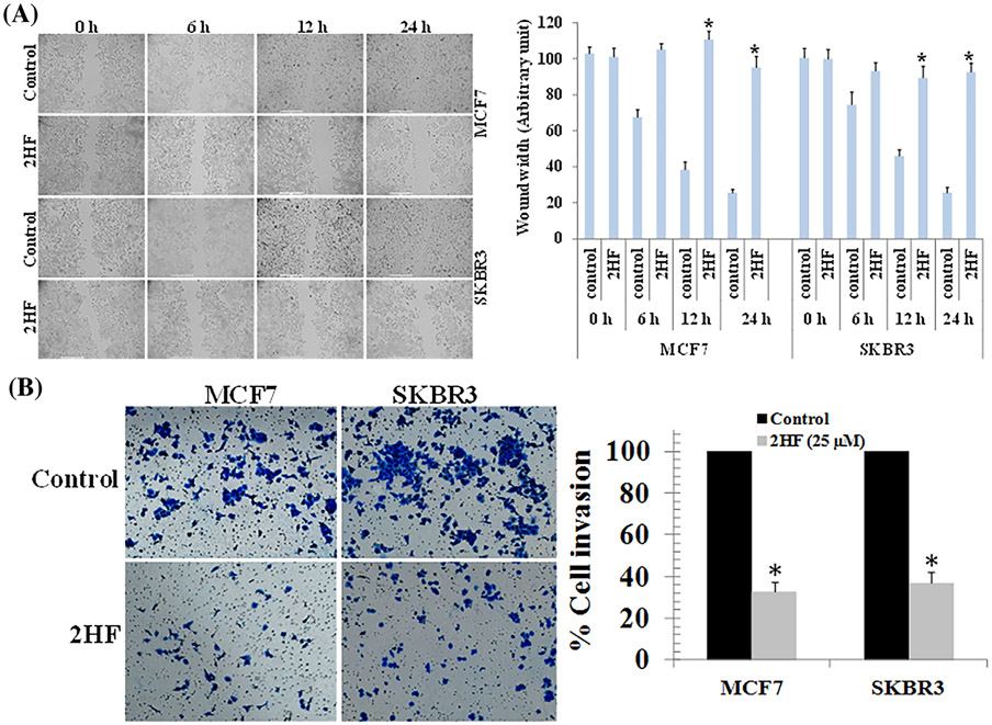
FIGURE 2.
Effects of 2HF on the migration and invasion of breast cancer cell lines. (A) MCF7 and SKBR3 BC cells were grown to 90% confluency in cell culture dishes. A scratch/wound was made in each dish. The cells were then treated with either vehicle or 2HF (25 μM). Images were taken at each time point for the respective control and treatment groups. The distance across the wound was measured for three replicate experiments and quantified as the wound width. (B) MCF7 and SKBR3 breast cancer cells were placed in the upper chamber of the transwell inserts (8 μm pore size) in serum-free medium, and then treated with either vehicle or 2HF (25 μM) for 24 h. The number of invaded cells were fixed with 4% paraformaldehyde and stained with 0.2% crystal violet. Invasive cells were then photographed under a light microscope at 200x. Quantification of invaded cells in 2HF treated cells is shown after normalizing to the percentage of invaded cells in control. Significantly different (*P < 0.01) compared with respective Controls by Student's t-test

