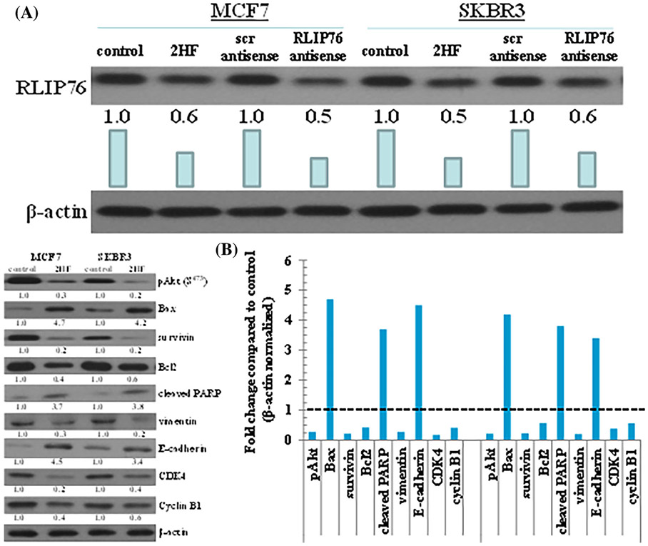
FIGURE 6.
Effects of 2HF on expression levels of protein in tumor tissues. Tumor tissue excised from control, 2HF-treated, scrambled (scr) antisense treated, and RLIP76-antisense treated MCF7 and SKBR3 xenograft mice were analyzed for changes in expression levels of RLIP76 (A), and Bax, Bcl2, cleaved PARP, survivin, CDK4, cyclin B1, E-cadherin, vimentin, and pAkt protein (B). β-actin was used as a loading control. Numbers below the blots represent the fold change in the levels of proteins as compared to Control as determined by densitometry. Bar diagram shows the quantification of respective Western blots. Dotted line represents no significant change as observed with control (panel B)

