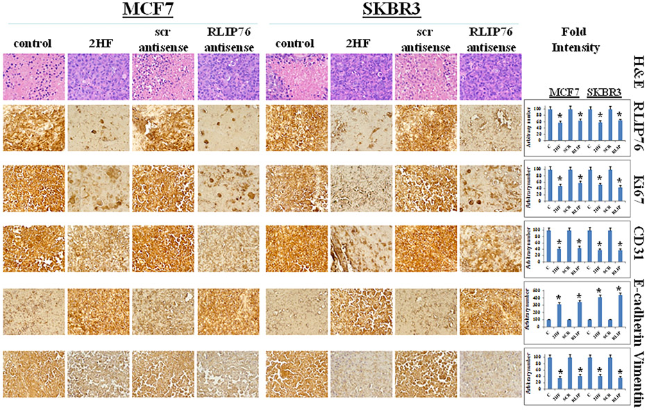
FIGURE 7.
Immunohistochemical analyses of RLIP76, and proliferation and angiogenesis markers in MCF7 and SKBR3 breast xenograft tumors. Tumor tissues from control, 2HF-treated, scrambled (scr) antisense treated, and RLIP76-antisense treated mice were used for Immunohistochemical analysis. Staining for H&E, RLIP76, Ki67, CD31, E-cadherin, and vimentin was performed. Photomicrographs at 40× magnification were acquired using Olympus DP 72 microscope. Percent staining was determined by measuring positive immuno-reactivity per unit area. The intensity of antigen staining was quantified by digital image analysis using Pro Plus software. Bars represent mean ± S.E. (n = 5). One representative image for each treatment group is shown. *Statistical significance of difference was determined by two-tailed Student's t-test; P < 0.01, 2HF and RLIP76-antisense treated compared with respective controls

