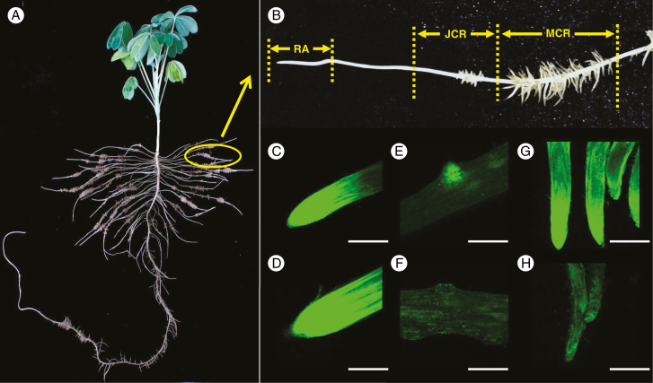
Fig. 1.
Flavonoid fluorescence in roots of white lupin grown under P-sufficient and P-deficient conditions. Plants were grown in hydroponic solution with 75 μm P (+P) or 0 μm P (−P) for 21 d. Root segments were stained with DPBA. Green fluorescence was detected by confocal laser scanning microscopy, which shows accumulation of flavonoids. (A) White lupin plant grown under −P conditions formed a large number of cluster roots. (B) Cluster roots were separated into root apex (RA), juvenile cluster roots (JCR) and mature cluster roots (MCR). (C–H) Images of flavonoid fluorescence in (C) −P root apex, (D) +P root apex, (E) −P juvenile cluster root, (F) +P juvenile cluster root, (G) −P mature cluster root and (H) +P mature cluster root. Scale bar = 1.0 mm.

