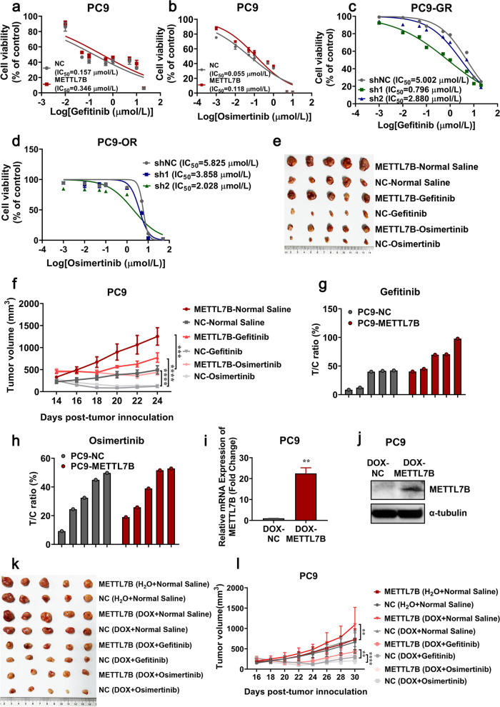
Fig. 2.
METTL7B induced resistance to TKIs in LUAD cells. a and b FLAG-NC and FLAG-METTL7B was stably transfected into TKIs-sensitive PC9 cell, and the cell viabilities were evaluated to measure IC50 of TKIs after treatment with different concentrations of gefitinib (a) and osimertinib (b) for 72 h. c and d shMETTL7B were stably transfected into gefitinib-resistant PC9-GR and osimertinib-resistant PC9-OR cells, and the cell viabilities were evaluated to measure IC50 of TKIs after incubation with different concentrations of gefitinib (c) and osimertinib (d) for 72 h. e–h PC9-FLAG-METTL7B or FLAG-NC were injected into the flank of BALB/c nude mice to form subcutaneous tumors. The tumors were treated with gefitinib and osimertinib every 2 d and the results were shown in tumor growth (e–f) and T/C ratio analysis (g-h) (n = 5 mice per group). T/C% = TRTV/CRTV × 100%; TRTV, relative tumor volume after drug treatment; CRTV, relative tumor volume of vehicle control. i-j PC9-pInducer-METTL7B[Tet-on] cells were treated with or without DOX (1 μM) for 72 h. The expression levels of METTL7B were assayed by qRT-PCR (i) and Western blot (j). k-l PC9-pInducer-METTL7B[Tet-on] cells were injected into the flank of BALB/c nude mice to form subcutaneous tumors. The CDX-bearing mice (n = 5 mice per group) were treated with vehicle (normal saline), gefitinib (30 mg/kg, qd, po) or osimertinib (30 mg/kg, qd, po) and DOX (1.5 mg/mL). The growth of tumors was monitored every 2d. **P < 0.01, ***P < 0.001 and ****P < 0.0001

