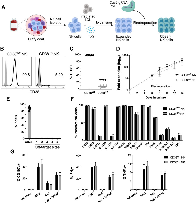
Figure 1.
Highly efficient method for CD38 KO in ex vivo expanded NK cells. (A) Ex vivo expansion and CRISPR/Cas9 gene editing protocol. NK cells were isolated from healthy donor PBMCs and cocultured with irradiated LCL feeder cells and IL-2 for 1 week. Expanded NK cells were electroporated using precomplexed Cas9 and CD38-targeting gRNA. Edited NK cells were then expanded further in IL-2 containing media and analyzed for KO efficiency, phenotype, and function 7 days after CRISPR/Cas9 editing. CD38WT NK cells were unmanipulated. (B) Representative histograms of relative expression of CD38 in CD38WT and CD38KO NK cells from one NK cell donor. (C) Pooled data showing relative expression of CD38 in CD38WT and CD38KO NK cells (n=8 donors). (D) Ex vivo fold expansion of CD38KO NK cells compared with unedited CD38WT NK cells (n=8 donors). (E) Assessment of off-target activity of the CD38-targeting gRNA/Cas9 RNP, shown are percentage of indels detected in CD38KO NK cells at the CD38 on-target site as well as five in-silico-predicted off-target sites, as analyzed using the on-line ICE tool (n=5 donors). (F) Expression of a panel of NK cell surface markers examined by flow cytometry in CD38KO and CD38WT NK cells (n=6 donors). (G) NK cell degranulation (measured by CD107a expression), and intracellular IFN-γ and TNF-α expression examined by flow cytometry of CD38KO NK cells compared with CD38WT NK cells following coculture with K562 cells and Raji cells with and without rituximab (RITUX) (n=6 donors). Statistics determined with the Student’s t-test, two tailed, ****p<0.0001. Differences between CD38WT and CD38KO NK cells in panels F and G were not significant. ICE, inference of CRISPR Edits; KO, knockout; WT, wild type; NK, natural killer; PBMC, peripheral blood mononuclear cell; LCL, lymphoblastoid cell line.

