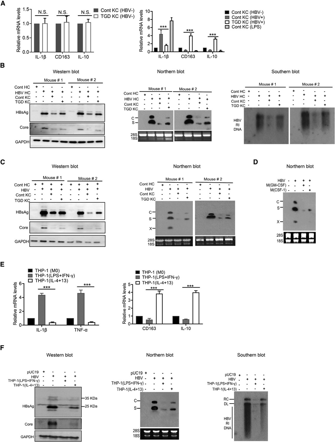
Figure 1. M1- and M2-like macrophages displayed different HBV-suppressive activities.

(A) Relative levels of IL-1β, CD163, and IL-10 mRNAs in KCs isolated from control (Cont) or TGD mice as quantified by qRT-PCR. Left panel, KCs without co-culturing with hepatocytes; right panel, KCs co-cultured with HBV-positive hepatocytes. In the right panel, KCs from control mice without co-culturing and those treated with LPSs were also used as the controls. N.S., statistically not significant; ***p < 0.001.
(B) KCs isolated from control and TGD mice were analyzed for their effects on HBV replication in hepatocytes isolated from HBV transgenic mice (HBV HCs) after 3 days of co-culturing. Left panel, western blot of HBV proteins; middle panel, northern blot for HBV RNAs; and right panel, Southern blot of HBV RI DNA. Hepatocytes isolated from control mice (Cont HCs) were used as the negative control. Note that the precore protein was not always visible in the western blot when the core protein was analyzed. In the middle panel, C and S are HBV C and S gene transcripts, respectively, and 28S and 18S rRNAs were used as the loading control.
(C) KCs isolated from control and TGD mice were analyzed for their effects on HBV replication in hepatocytes isolated from mice 1 day after hydrodynamic injection of the 1.3-mer HBV genomic DNA. Hepatocytes were co-cultured with KCs for 3 days and lysed for analysis of HBV proteins by western blot (left panel) or RNAs by northern blot (right panel). The X gene transcript was also detected in the northern blot.
(D) M(GM-CSF) or M(CSF-1) were treated with LPSs for 3 h and then co-cultured with HBV-infected HHs for 14 days. HHs were then lysed for northern blot analysis of HBV RNAs.
(E) THP-1(M0), THP-1(LPS+IFN-γ), and THP-1(IL-4+13) were lysed for analysis of IL-1β, TNF-α, IL-163, and IL-10 RNAs by qRT-PCR.
(F) Huh7 cells were transfected with the control vector pUC19 or the 1.3-mer HBV genomic DNA and, 6 h after DNA transfection, co-cultured with THP-1(LPS+IFN-γ) or THP-1(IL-4+13) for 2 days. Huh7 cells were then lysed for analysis of HBV proteins (left panel), HBV RNA (middle panel), and HBV DNA (right panel). RC, relaxed circular DNA of HBV; DL, double-stranded linear form of HBV DNA.
See also Figure S1.
