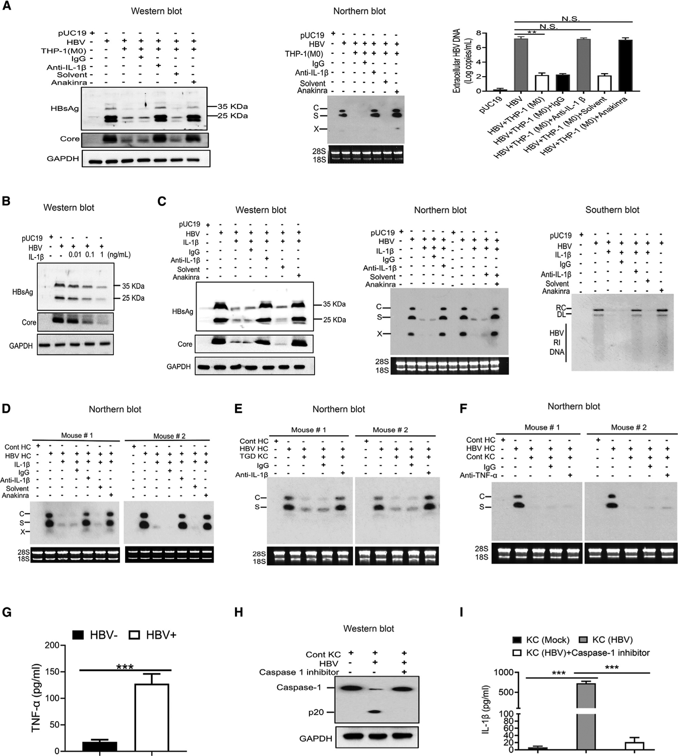
Figure 3. Suppression of HBV replication by IL-1β.

(A) Huh7 cells that had been transfected with pUC19 or pHBV1.3mer were co-cultured with THP-1 macrophages, either in the presence of a control immunoglobulin G (IgG) or the anti-IL-1β antibody (R&D) or in the presence of anakinra or its solvent (i.e., water). Huh7 cells were lysed 2 days after co-culturing for analysis of HBV proteins or HBV RNAs (middle panel). The virion-associated HBV DNA in the incubation media was also quantified by qPCR (right panel). N.S., statistically not significant; **p < 0.01.
(B) Huh7 cells, 2 days after transfection with pHBV1.3mer, were treated with 0.01, 0.1, or 1 ng/mL of IL-1β for 1 day and then lysed for western blot analysis of HBV proteins. Huh7 cells transfected with pUC19 were used as the control.
(C) Huh7 cells transfected with pHBV1.3mer were treated with IL-1β (1 ng/mL) as mentioned above in the presence of control IgG, anti-IL-1β antibody, or anakinra. Cells were then lysed for analysis of HBV proteins (left panel), RNAs (middle panel), and DNA (right panel).
(D) Tg05 HBV transgenic mice were peritoneally injected with IL-1β (200 ng/mouse) with or without the co-injection of control IgG, anti-IL-1β antibody, or anakinra. Hepatocytes were isolated from mice 72 h after injection for the analysis of HBV RNAs.
(E) KCs isolated from TGD mice were co-cultured with hepatocytes isolated from HBV transgenic mice in the presence of a control IgG or the anti-IL-1β antibody. Hepatocytes were lysed 72 h later for analysis of HBV RNAs.
(F) The experiment was conducted the same way as in (E), except that the anti-IL1β antibody was replaced by the anti-TNF-α antibody.
(G) The level of TNF-α in the incubation media of KCs in (F) with (HBV+) or without HBV (HBV−) stimulation was measured by ELISA.
(H) KCs isolated from control mice and co-cultured with HBV-positive hepatocytes in the absence or presence of the caspase 1 inhibitor ex vivo for 72 h were lysed for western blot analysis of caspase-1.
(I) The experiment was conducted as in (H), except that the incubation media were harvested for quantification of IL-1β by ELISA.
See also Figure S3.
