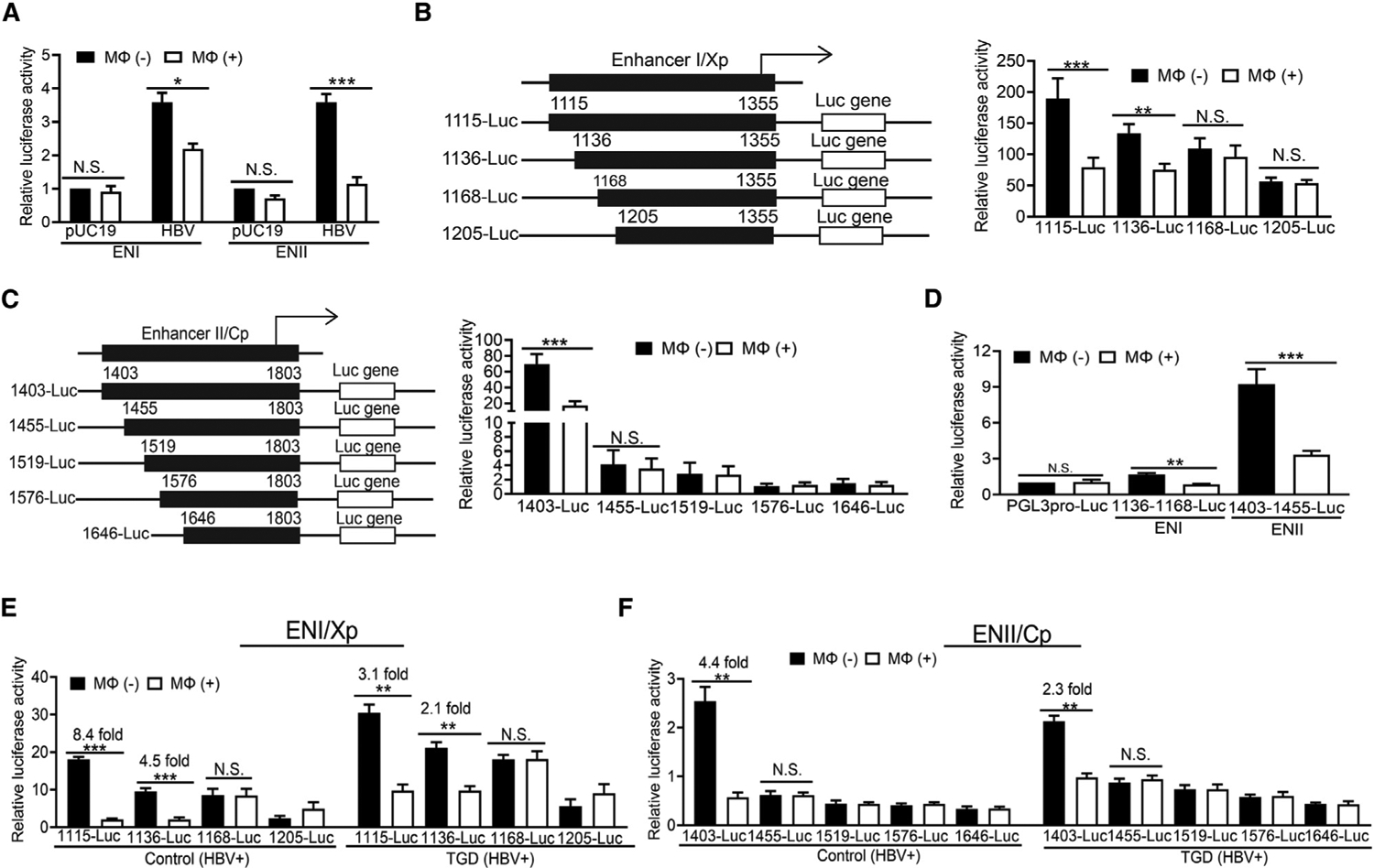
Figure 4. Suppression of HBV ENI and ENII enhancers by macrophages.

(A) HBV ENI or ENII enhancer reporter construct was co-transfected with pUC19 or pHBV1.3mer into Huh7 cells. The renilla luciferase reporter pRL-TK was also used in the co-transfection to monitor the transfection efficiency. Cells with (Mφ+) or without (Mφ–) co-culturing with THP-1 macrophages for 2 days were then lysed for the measurement of luciferase activities.
(B) The ENI reporter constructs with different deletions are illustrated to the left. These reporter constructs were transfected into HepAD38 cells, and the relative luciferase activities of the reporter constructs in the absence (–) or presence (+) of macrophages are shown in the chart to the right.
(C) The ENII reporter constructs with different deletions are illustrated to the left. The studies were conducted the same way as described in (B).
(D) THP-1 macrophages reduced the activities of 1,136–1,168-luciferase (Luc) and 1,403–1,455-Luc reporter constructs in HepAD38 cells.
(E) Hepatocytes isolated from control or TGD mice that had been injected with 20 μg pHBV1.3mer were transfected with the ENI reporter constructs ex vivo. These hepatocytes, with (+) or without (−) the subsequent co-culturing with their paired KCs isolated from the same mice, were then lysed for analysis of the reporter activities.
(F) The study was conducted the same way as in (E), with the exception that ENII reporter constructs were analyzed. The results represent the mean ± SEM of three independent experiments. N.S., not significant; *p < 0.05; **p < 0.01; ***p < 0.001.
See also Figure S4.
