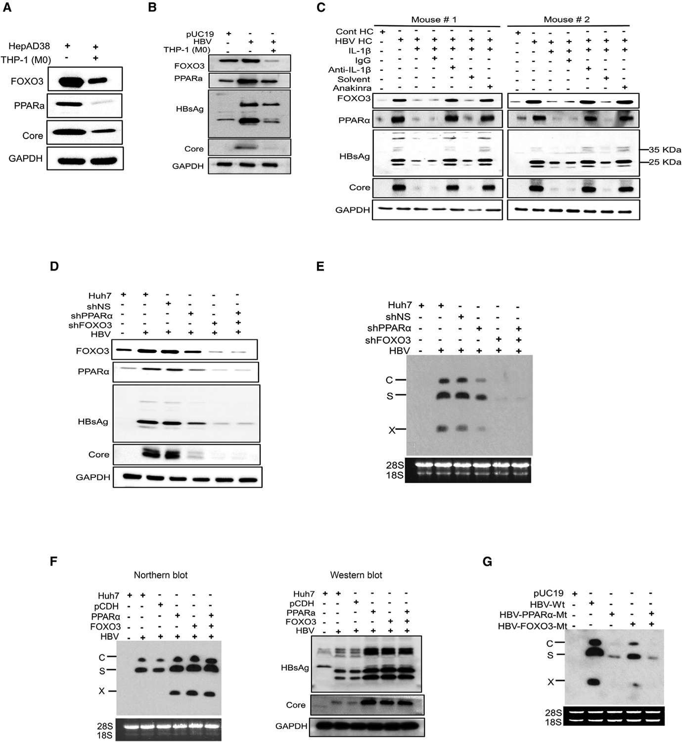
Figure 6. Analysis of the effects of PPARα and FOXO3 on HBV gene expression.

(A) HepAD38 cells with replicating HBV were co-cultured with THP-1 macrophages for 2 days and lysed for western blot analysis of FOXO3, PPARα, and HBV core protein. GAPDH was used as the loading control.
(B) Huh7 cells transfected with the pHBV1.3mer with or without co-culturing with THP-1 macrophages were lysed for western blot analysis of FOXO3, PPARα, HBsAg, and the HBV core protein. Huh7 cells transfected with pUC19 was used as the control.
(C) Tg05 HBV transgenic mice were peritoneally injected with IL-1β (200 ng/mouse) with or without the co-injection of control IgG, anti-IL-1β antibody, or anakinra. Hepatocytes were isolated from mice 24 h later and lysed for western blot analysis.
(D) Huh7 cells transduced with the lentiviral vector that expressed a scrambled shRNA (shNS), PPARα shRNA (shPPARα), or FOXO3 shRNA (shFOXO3) were transfected with the pHBV1.3mer. Cells were lysed 48 h later for western blot analysis.
(E) Experiments were conducted the same way as in (D), followed by northern blot analysis of HBV RNAs.
(F) Huh7 cells were transduced with the lentiviral vector that expressed either PPARα, FOXO3, or both and then transfected with pHBV1.3mer. Cells were lysed 48 h after transfection for analysis of HBV RNAs and proteins. The control lentiviral vector was also used to serve as the control.
(G) Huh7 cells were transfected with pHBV1.3mer or its mutants that contained mutations in the binding site of either PPARα, FOXO3, or both and lysed 48 h later for northern blot analysis of HBV RNAs.
See also Figure S5.
