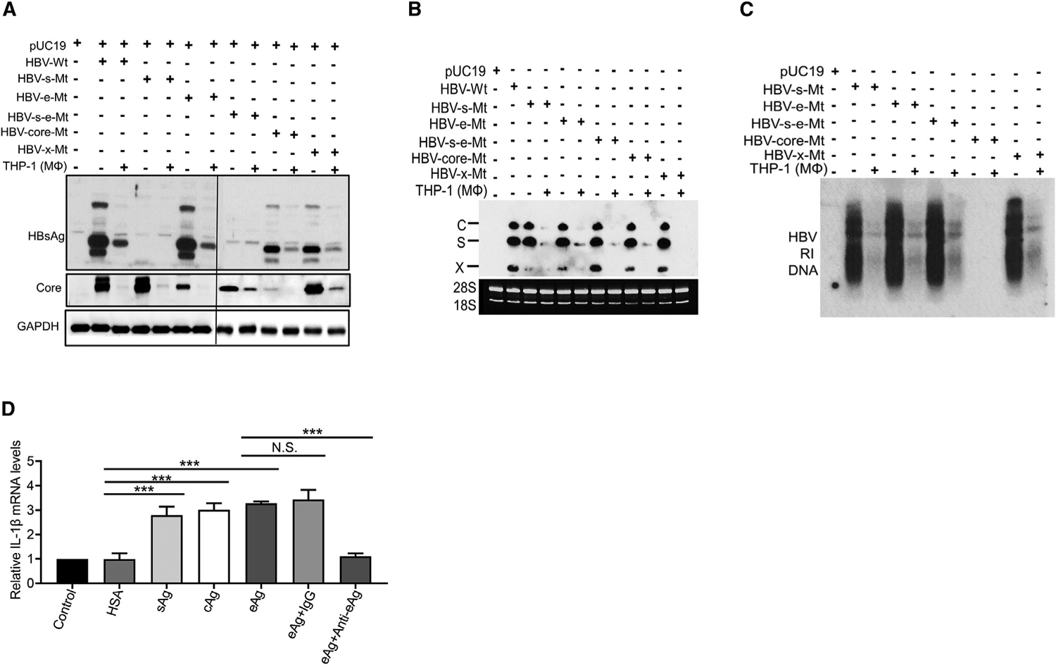
Figure 7. Analysis of the response of THP-1 macrophages to various HBV proteins.

(A–C) Huh7 cells transfected with pUC19 or the HBV genomic DNA with various mutations, and with or without subsequent co-culturing with THP-1 macrophages, were lysed and analyzed by western blot (A), northern blot (B), or Southern blot (C).
(D) THP-1 macrophages not treated (NT) or treated with HSA, HBsAg, HBV core protein, or HBeAg were lysed for qRT-PCR analysis of the IL-1β RNA. The IL-1β RNA level of cells not treated with recombinant proteins was arbitrarily defined as 1. In the HBeAg studies, the addition of the anti-HBeAg antibody, but not the control antibody, abolished the induction of IL-1β by HBeAg in THP-1 macrophages. The results represented the mean ± SEM of three independent experiments.
N.S., not significant; ***p < 0.001.
See also Figure S6.
