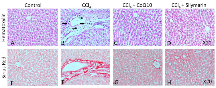
Figure 5.
Effect of CCl4 and supplementation with CoQ10 and silymarin on liver tissue. Hemtoxylin-eosin staining shows the cellular morphology in control (A), CCl4 (B), CCl4 + CoQ10 (C), and CCl4 + silymarin (D) liver of ovariectomized rats. Arrows indicate infiltrating cells. Sirius Red staining depicts fibrosis in control (E), CCl4 (F), CCl4 + CoQ10 (G), and CCl4 + silymarin (H) liver of ovariectomized rats. 20× magnification.

