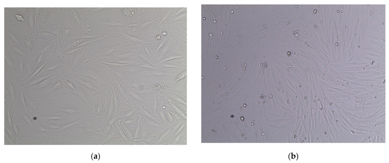
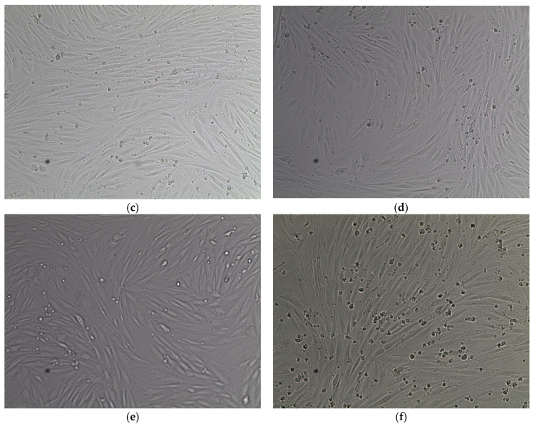
Figure 1.
Images depicting L6 skeletal muscle myoblasts from rats, differentiated into L6 skeletal muscle myotubes (passage 7). (a) Myoblasts obtained on day 3 of the tissue culture in 10% FBS Ham F-10 media (10×). (b) Cells obtained on day 4 of the tissue culture in 6% horse serum Ham F-10 media (10×). (c) Myotubes obtained on day 5 of the tissue culture in 2% horse serum Ham F-10 media (10×). (d) Myotubes obtained after 4 h of tissue culture in 2% delipidated serum Ham F-10 media (10×). (e) Myotubes obtained after one hour of cell starvation in only Ham F-10 media (10×). (f) Myotubes obtained after 19 h of cell starvation in only Ham F-10 media (10×).


