. 2022 Jan 14;4(1):e210063. doi: 10.1148/rycan.210063

Figure 1:

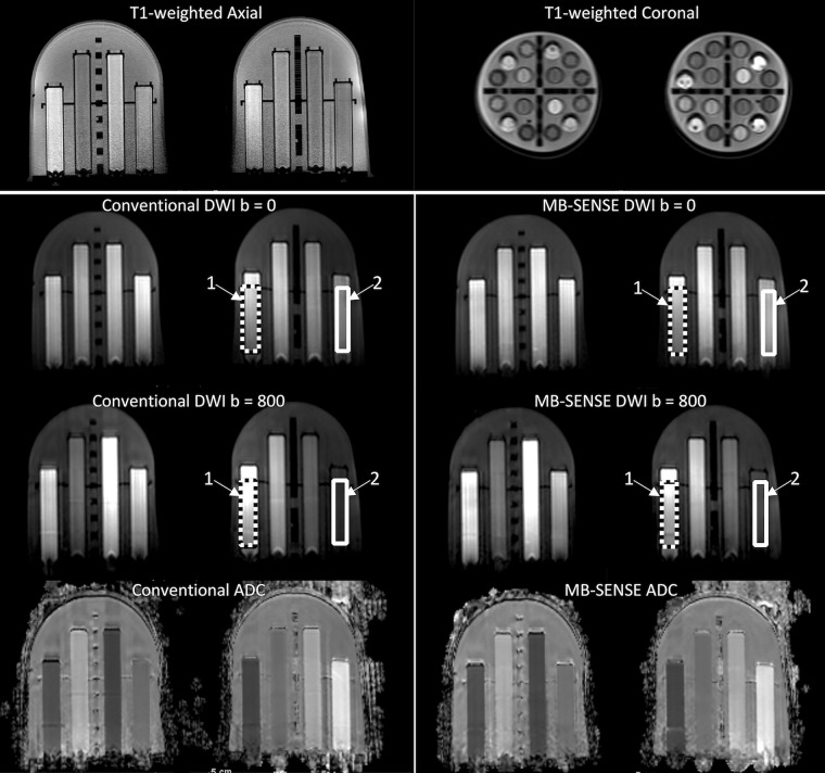
Images in a bilateral breast diffusion-weighted imaging (DWI) phantom. Shown are T1-weighted axial and coronal cross-section views (top) and representative images obtained at DWI (b = 0 and 800 sec/mm2) that depict the tumor mimic (arrow 1) and the normal tissue mimic (arrow 2) vials in the left breast used for quantitation of the ADC, contrast-to-noise ratio, and signal index. Corresponding regions of interest used for quantitation are shown for tumor tissue (dotted line) and normal tissue (solid line) mimics. ADC = apparent diffusion coefficient, MB = multiband, SENSE = sensitivity encoding.

Images in a bilateral breast diffusion-weighted imaging (DWI) phantom. Shown are T1-weighted axial and coronal cross-section views (top) and representative images obtained at DWI (b = 0 and 800 sec/mm2) that depict the tumor mimic (arrow 1) and the normal tissue mimic (arrow 2) vials in the left breast used for quantitation of the ADC, contrast-to-noise ratio, and signal index. Corresponding regions of interest used for quantitation are shown for tumor tissue (dotted line) and normal tissue (solid line) mimics. ADC = apparent diffusion coefficient, MB = multiband, SENSE = sensitivity encoding.